RNA N6-methyladenosine modulates endothelial atherogenic responses to disturbed flow in mice

  1. Bochuan Li
  2. Ting Zhang
  3. Mengxia Liu
  4. Zhen Cui
  5. Yanhong Zhang
  6. Mingming Liu
  7. Yanan Liu
  8. Yongqiao Sun
  9. Mengqi Li
  10. Yikui Tian
  11. Ying Yang  Is a corresponding author
  12. Hongfeng Jiang  Is a corresponding author
  13. Degang Liang  Is a corresponding author
  1. Tianjin Key Laboratory of Metabolic Diseases, Key Laboratory of Immune Microenvironment and Disease (Ministry of Education), Collaborative Innovation Center of Tianjin for Medical Epigenetics and Department of Physiology and Pathophysiology, Department of Cardiovascular Surgery, Tianjin Medical University General Hospital, Tianjin Medical University, China
  2. CAS Key Laboratory of Genomic and Precision Medicine, Collaborative Innovation Center of Genetics and Development, College of Future Technology, Beijing Institute of Genomics, Chinese Academy of Sciences, China
  3. China National Center for Bioinformation, China
  4. University of Chinese Academy of Sciences, China
  5. Key Laboratory of Remodeling-Related Cardiovascular Diseases (Ministry of Education), Beijing Collaborative Innovation Center for Cardiovascular Disorders, Beijing Institute of Heart Lung and Blood Vessel Diseases, Beijing Anzhen Hospital, Capital Medical University, China

Abstract

Atherosclerosis preferentially occurs in atheroprone vasculature where human umbilical vein endothelial cells are exposed to disturbed flow. Disturbed flow is associated with vascular inflammation and focal distribution. Recent studies have revealed the involvement of epigenetic regulation in atherosclerosis progression. N6-methyladenosine (m6A) is the most prevalent internal modification of eukaryotic mRNA, but its function in endothelial atherogenic progression remains unclear. Here, we show that m6A mediates the epidermal growth factor receptor (EGFR) signaling pathway during EC activation to regulate the atherosclerotic process. Oscillatory stress (OS) reduced the expression of methyltransferase like 3 (METTL3), the primary m6A methyltransferase. Through m6A sequencing and functional studies, we determined that m6A mediates the mRNA decay of the vascular pathophysiology gene EGFR which leads to EC dysfunction. m6A modification of the EGFR 3’ untranslated regions (3’UTR) accelerated its mRNA degradation. Double mutation of the EGFR 3’UTR abolished METTL3-induced luciferase activity. Adenovirus-mediated METTL3 overexpression significantly reduced EGFR activation and endothelial dysfunction in the presence of OS. Furthermore, thrombospondin-1 (TSP-1), an EGFR ligand, was specifically expressed in atheroprone regions without being affected by METTL3. Inhibition of the TSP-1/EGFR axis by using shRNA and AG1478 significantly ameliorated atherogenesis. Overall, our study revealed that METTL3 alleviates endothelial atherogenic progression through m6A-dependent stabilization of EGFR mRNA, highlighting the important role of RNA transcriptomics in atherosclerosis regulation.

Editor's evaluation

Methylation of adenine residues in mRNA has been shown to be a regulator of many factors in heath and disease. In these studies, the authors present data that this modification of the mRNA for EGFR (epidermal growth factor receptor) through down regulation of the methylating enzyme METTL3 by shear stress is a contributor to vascular pathology in a model with some features of accelerated atherosclerosis.

https://doi.org/10.7554/eLife.69906.sa0

Introduction

N6-methyladenosine (m6A) is the most prevalent post-transcriptional modification of eukaryotic mRNAs (Roundtree et al., 2017). This modification is reversible and is catalyzed by a multicomponent methyltransferase complex consisting of various methyltransferases including methyltransferase like 3 (METTL3), METTL14, Wilms tumor 1-associated protein (WTAP), and KIAA1429 (Virilizer), and is erased by demethylases such as fat mass and obesity-associated protein or α-ketoglutarate-dependent dioxygenase alk B homolog 5 (Shi et al., 2019; Huang et al., 2021). As previously reported, m6A on mRNAs plays an important role in regulating cellular processes, including RNA stability, translation efficiency, RNA secondary structure, subcellular localization, alternative polyadenylation, and splicing (Liu et al., 2015; Wang et al., 2015; Zaccara et al., 2019). In the methyltransferase complex, METTL14 functions as the target recognition subunit by binding to RNA and then recruits METTL3 to catalyze m6A formation. Wilms tumor 1-associated protein (WTAP) is a regulatory subunit required for the accumulation of METTL3 and METTL14 into nuclear speckles (Ping et al., 2014). KIAA1429 guides region-selective m6A methylation (Yue et al., 2018). As the core methyltransferase subunit, METTL3 has been demonstrated to modulate key physiological processes, including spermatogenesis (Xu et al., 2017), cell reprogramming (Chen et al., 2015), and embryonic stem cell chromatin modification (Liu et al., 2021; Xu et al., 2021). However, its function in human cardiovascular disease (CVD) remains elusive.

Atherosclerosis, resulted from endothelial dysregulation in the arterial wall (Geovanini and Libby, 2018), is the leading cause of CVD resulting in high rate of mortality in the population. Atherosclerosis preferentially develops at branches and curvatures in the arterial tree where flow is disturbed (Davies, 2009). Disturbed flow pattern increases inflammatory response in ECs, including the expression of intercellular adhesion molecule 1 and vascular adhesion molecule 1 (VCAM-1) (Humphrey et al., 2014). Recent studies have reported that oscillatory stress (OS) could modulate atherosclerosis development by inducing the expression of DNA methyltransferases (Dunn et al., 2014) and histone modifications (Hastings et al., 2007), indicating the involvement of epigenetic mechanisms in the regulation of atherogenesis. In this study, we focused on exploring the function of RNA m6A modification under OS in ECs, which may provide a better understanding of atherogenesis.

Numerous studies have shown that thrombospondin-1 (TSP-1, encoded by THBS1) is a shear-sensitive protein important for the regulation of vascular remodeling (Ni et al., 2010). Notably, TSP-1 can induce dysregulated blood flow, impaired vessel dilation, and increased vascular tone Csányi et al., 2012 followed by arterial stiffening (Kim et al., 2017). In addition, Thbs 1 deficiency prevented lesion formation in Apoe-/- mice (Ganguly et al., 2017). As a receptor of TSP-1, epidermal growth factor receptor (EGFR) has been reported to be involved in vascular pathophysiology and pathogenesis of atherosclerosis in macrophages (Wang et al., 2017). Moreover, EGFR-selective tyrphostin, AG1478, can reverse the phosphorylation of EGFR tyrosine-1068 induced by TSP-1 activation (Liu et al., 2009), indicating the complicated regulation between TSP-1 and EGFR with respect to vascular function.

In this study, we generated tamoxifen-inducible endothelial-specific Mettl3-deficient (EC-Mettl3KO) mice to investigate the regulation of Mettl3-mediated m6A on atherogenesis. Our data revealed that downregulation of METTL3 and hypomethylation mediate OS-induced endothelial dysfunction and atherogenesis both in vivo and in vitro, indicating that the THBS1/EGFR axis is a key regulatory target of METTL3-dependent EC activation. These results illuminate a critical mechanism of m6A modification in regulating atherosclerosis.

Results

METTL3 is decreased in atheroprone regions

To explore the functions of m6A modification under OS, we first detected m6A level changes in response to OS (0.5 ± 4 dyn/cm2, 1 Hz) for 6 hr in human umbilical vein ECs (HUVECs) through UHPLC-MRM-MS (ultra-high-performance liquid chromatography-triple quadrupole mass spectrometry coupled with multiple-reaction monitoring) analysis. The results showed that OS significantly decreased m6A modification in HUVECs (Figure 1A). To further investigate the effects of OS on the pattern of m6A modulators, we examined methyltransferase components including METTL3, METTL14, METTL16, WTAP, and Virilizer. Western blot analysis detected a significantly decrease of METTL3 and Virillizer after OS stimulation, without affecting other m6A modulators (Figure 1B–E). Furthermore, to analyze the m6A modulators in atheroprone (aortic arch [AA]) and atheroprotective (thoracic aorta [TA]) regions, we performed western blot analysis and found that METTL3 and Virilizer were significantly decreased in AA tissue lysates than in TA region, whereas METTL14 and WTAP were not changed (Figure 1—figure supplement 1A and B). Consistently, in outer curvature of AA and TA, where blood flow is laminar, METTL3 was highly expressed in both regions but only displayed specific nuclear localization in outer curvature of AA. Conversely, in the inner curvature and bifurcation of AA, where blood flow is disturbed, METTL3 was dramatically decreased (Figure 1F and G; Figure 1—figure supplement 1C). Next, to further test the expression of METTL3, we used Apoe-/- mice with partial ligation and performed carotid Doppler ultrasonography to verify disturbed flow induction in the partially ligated left common carotid artery (LCA) at 7th and 14th days after the procedure compared with stable flow in those at day 0 (Figure 1—figure supplement 1D). Consistent with the AA, as compared with right common carotid artery (RCA), METTL3 was weakly expressed in LCA after 1 and 2 weeks (Figure 1—figure supplement 1E and F). Collectively, METTL3 is decreased in atheroprone regions.

Figure 1 with 1 supplement see all
Methyltransferase like 3 (METTL3)-dependent N6-methyladenosine (m6A) methylation is decreased in atheroprone regions.

Human umbilical vein endothelial cells (HUVECs) and mouse aortic endothelial cells (mAECs) were exposed to OS (0.5 ± 4 dyn/cm2) for 6 hr. Cells with static treatment (ST) were a control. (A) Ultra-high-performance liquid chromatography-triple quadrupole mass spectrometry coupled with multiple-reaction monitoring analysis of m6A levels in mRNAs extracted from HUVECs exposed to ST and OS. Data are shown as the mean ± SEM, *p<0.05, NS, not significant (Student’s t test). n = 3. (B–E) Western blot analysis of METTL3, METTL14, METTL16, Wilms tumor 1-associated protein, and Virillizer expression in HUVECs (B–C) and mAECs (D–E) response to ST and OS. Data are mean ± SEM, *p<0.05 (Student’s t test). n = 6. (F) Aortas from 6- to 8-week-old Apoe-/- mice underwent immunofluorescence staining for indicated proteins. AA inner, inner curvature of aortic arch; AA outer, outer curvature of aortic arch; TA, thoracic aorta. Scale bar, 20 μm. (G) Quantification of protein expression in (F). Data are mean ± SEM, *p<0.05 (one-way ANOVA with Bonferroni multiple comparison post-test). n = 6.

Figure 1—source data 1

METTL3-dependent m6A methylation is decreased in atheroprone regions.

https://cdn.elifesciences.org/articles/69906/elife-69906-fig1-data1-v2.zip

Endothelial activation arises in EC-specific METTL3-deficient mice

Next, to clarify whether METTL3 deficiency induces endothelial dysfunction, we crossed tamoxifen-inducible endothelial-specific Mettl3-deficient (EC-Mettl3KO) mice (Figure 2—figure supplement 1A and B). METTL3 was used as a marker of knockout efficiency. The protein levels of METTL3 were decreased in AA intima tissue lysates compared to TA in Mettl3flox/flox mice. The level of the EC activation marker VCAM-1 was significantly increased in AA intima tissue lysates compared to TA intima lysates in both Mettl3flox/flox mice and EC-Mettl3KO mice (Figure 2A–C). Next, we used EC-Mettl3KO mice and Mettl3flox/flox mice with partial ligation to further test the expression of METTL3. En face immunofluorescence staining of LCA revealed reduced protein levels of METTL3 and enhanced levels of VCAM-1 in EC-Mettl3KO mice compared to RCA 2 weeks after ligation (Figure 2D and E). Overexpression of METTL3 in ECs by adeno-associated virus (AAV9-METTL3 OE) inhibited VCAM-1 expression induced by partial ligation in LCA endothelium (Figure 2F and G; Figure 2—figure supplement 1C and D). These results indicate that METTL3 depletion is associated with EC activation in response to OS.

Figure 2 with 1 supplement see all
Methyltransferase like 3 (METTL3) deficiency induces endothelial activation in atheroprone regions.

(A–C) Protein was extracted from the AA and TA of 8-week-old EC-Mettl3KO and Mettl3flox/flox mice. (A) Western blot analysis of the expression of METTL3, vascular adhesion molecule (VCAM-1), CD31, SMA (smooth muscle actin), and GAPDH in tissue lysates of AA and TA intima. AA, aortic arch; TA, thoracic aorta. (B–C) Quantification of protein expression in (A). Data are shown as the mean ± SEM, *p<0.05 (two-way ANOVA with Bonferroni multiple comparison post hoc test). Protein extracts of intima from three mice were pooled as one sample, n = 3. (D) EC-Mettl3KO and Mettl3flox/flox mice underwent partial ligation of the carotid artery for 2 weeks. En face immunofluorescence staining for the expression of VCAM-1 and METTL3 in ECs of the carotid artery of mice. Scale bar, 20 μm. (E) Quantification of the relative fluorescence intensity of VCAM-1 and METTL3. Data are shown as the mean ± SEM, *p<0.05 (two-way ANOVA with Bonferroni multiple comparison post hoc test). n = 6 mice. (F) Male mice underwent partial ligation of the carotid artery. During ligation, carotid arteries were infused with the indicated adeno-associated virus. Immunofluorescence staining of VCAM-1 and vWF in ECs of the RCA and LCA of mice. RCA, right carotid artery; LCA, left carotid artery. Scale bar, 20 μm. (G) Quantification of the relative fluorescence intensity of VCAM-1. Data are shown as the mean ± SEM, *p<0.05 (one-way ANOVA with Bonferroni multiple comparison post hoc test). n = 5 mice.

Figure 2—source data 1

Mettl3 deficiency induces endothelial activation in atheroprone regions.

https://cdn.elifesciences.org/articles/69906/elife-69906-fig2-data1-v2.zip

OS-abolished m6A prevents EGFR mRNA degradation

As a core subunit of the m6A methyltransferase complex, the downregulation of METTL3 expression in response to OS suggests the potential regulation of m6A modification. First, we conducted m6A-specific methylated RNA immunoprecipitation combined with high-throughput sequencing (MeRIP-seq) to compare the landscape of m6A in static treatment (ST) and OS. We identified 10,515 and 10,580 m6A peaks in ST and OS, respectively, all of which were enriched in coding regions, 3’ untranslated regions (3’UTRs), and near stop codons (Figure 3A; Figure 3—figure supplement 1A). Interestingly, we found that the m6A motifs identified in OS and ST were significantly enriched in GGACU (Figure 3B). Furthermore, we identified thousands of dysregulated m6A peaks induced by OS (Figure 3—figure supplement 1B; Supplementary file 1), and genes with downregulated m6A peaks were significantly enriched in transcription regulation- and cell-cell and membrane adhesion-related pathways (Figure 3—figure supplement 1C, upper panel). Overall, we found that METTL3 may mediate the dynamic change in m6A landscapes between ST and OS.

Figure 3 with 1 supplement see all
Oscillatory stress (OS)-abolished N6-methyladenosine (m6A) prevents epidermal growth factor receptor (EGFR) mRNA degradation.

(A) Distribution of m6A peaks along the 5’ untranslated regions (5’UTR), CDS (coding sequence), and 3’UTR regions of mRNA in static treatment (ST) and OS. (B) m6A motif identified from human umbilical vein endothelial cells (HUVECs) under ST and OS treatments. (C) Integrative genomics viewer tracks displaying the results of IP vs. input read distributions in EGFR 3’UTR mRNA of HUVECs under ST and OS treatments. (D) qPCR analysis of EGFR mRNA levels in ST and OS. Data are shown as the mean ± SEM, *p<0.05 (Student’s t test). n = 6. (E) MeRIP-qPCR detection of m6A enrichment on EGFR mRNA in ST and OS. Data are shown as the mean ± SEM, *p<0.05 (Student’s t test). n = 5. (F–H) qPCR analysis showing delayed EGFR mRNA degradation upon methyltransferase like 3 (Mettl3)-overexpression (F); si-Mettl3 (G); and OS treatment (H). HUVECs were treated with actinomycin D for 4 and 6 hr. Data are shown as the mean ± SEM, *p<0.05 (two-way ANOVA with Bonferroni multiple comparison post hoc test). n = 6. (I) Schematic representation of the mutated (RRACH to RRTCH) 3’ UTR of EGFR plasmids. (J) Relative activity of the wild-type or mutant EGFR 3’UTR firefly luciferase reporter in K293 cells treated with green fluorescent protein (GFP)- or GFP-Mettl3-overexpressing adenovirus. Data are shown as the mean ± SEM, *p<0.05, NS, not significant (Student’s t test). n = 8. RNA-seq and MeRIP-seq data generated in this study have been deposited to the Genome Sequence Archive in BIG Data Center under accession number PRJCA004746.

Figure 3—source data 1

OS-abolished m6A prevents EGFR mRNA degradation.

https://cdn.elifesciences.org/articles/69906/elife-69906-fig3-data1-v2.zip

As reported, m6A modification plays a very important role in regulating RNA abundance (Wang et al., 2014). To investigate the molecular mechanisms of m6A function in endothelial activation, RNA-seq was performed using ECs treated with ST and OS. We identified 547 consistently upregulated genes (Figure 3—figure supplement 1D-F) upon OS. Consistent with the MeRIP-seq data, these upregulated genes were also significantly enriched in transcription regulation- and cell adhesion-associated pathways (Figure 3—figure supplement 1G, right panel). Therefore, we hypothesized that METTL3 regulates the degradation of these genes in response to OS. Furthermore, we analyzed the upstream regulators of methylation-downregulated but expression-upregulated genes using IPA software. According to the IPA analysis, we identified EGFR as a potential key regulator participating in OS-enhanced gene ontology (GO) pathways (Figure 3—figure supplement 1D; Supplementary file 2), which was further confirmed by the reduced ratio of EGFR (IP/input) in OS compared to ST (Figure 3C). We also validated the significantly increased EGFR mRNA levels (Figure 3D) and decreased m6A enrichment in OS compared to ST by MeRIP-quantitative PCR (qPCR) (Figure 3E). To further test whether METTL3-mediated m6A regulates EGFR mRNA decay, we measured EGFR mRNA levels in ECs after treatment with the transcriptional inhibitor actinomycin D. Compared to the green fluorescent protein (GFP) control, METTL3 overexpression significantly decreased the remaining EGFR mRNA levels due to accelerated mRNA decay in the presence of functional m6A modification by overexpression of METTL3 (Figure 3F). As expected, EGFR mRNA showed a slower decay rate in response to METTL3 knockdown or OS treatment (Figure 3G and H). As we found that the 3’UTR of EGFR is a key region regulating m6A modification, we mutated two nearby potential m6A motifs AGACA and GGACT to AGTCA and GGTCT. Notably, the 3’UTR mutants did not respond to the decrease in luciferase activity caused by overexpression of METTL3 (Figure 3I and J). Collectively, these results suggest that OS-induced EC activation was mediated by m6A modification on EGFR.

TSP-1/EGFR pathway participates in EC inflammation induced by METTL3 inhibition in response to OS

EGFR ligands that specifically activate EGFR include epidermal growth factor (EGF), transforming growth factor α (TGF-α), and TSP-1 (Liu et al., 2009). TSP-1 is a multidomain protein that contains EGF-like repeats that indirectly activate EGFR and selected downstream signaling pathways (Garg et al., 2011; Liu et al., 2009). TSP-1 is activated in shear-mediated arterial stiffening (Kim et al., 2017). To study the function of the endothelial TSP-1/EGFR axis in EC activation, we first treated wildtype and METTL3-overexpressed ECs with or without OS. Overexpression of METTL3 abolished phosphorylation of EGFR (Tyr-1068), AKT, and ERK, as well as total EGFR and VCAM-1 levels in response to OS (Figure 4A and B; Figure 4—figure supplement 1A). Furthermore, TSP-1 was significantly increased at both the mRNA and protein levels under OS (Figure 4—figure supplement 1B). Supplementation with recombinant human TSP-1 didn’t change EGFR protein levels but enhanced EGFR phosphorylation which could be abolished by METTL3 overexpression (Figure 4C and D). Next, ECs were subjected to OS or METTL3 siRNA in the presence of the EGFR-selective tyrphostin AG1478, which blocked both OS and siMETTL3-mediated phosphorylation of EGFR, AKT, and ERK and VCAM-1 levels, had no effect on total EGFR expression (Figure 4E–H). The increased number of THP-1 (human myeloid leukemia mononuclear) cells adhering to HUVECs by OS or siMETTL3 was relieved in the presence of overexpression of METTL3 or AG1478 (Figure 4F and H; Figure 4—figure supplement 1C-D). These results suggest that METTL3 inhibits the transcriptional level of EGFR and downstream signaling events and cellular responses.

Figure 4 with 1 supplement see all
The thrombospondin-1/epidermal growth factor receptor (TSP-1/EGFR) pathway participates in EC inflammation induced by methyltransferase like 3 (METTL3) inhibition in response to oscillatory stress (OS).

(A) Western blot analysis of p-EGFR, EGFR, p-AKT, t-AKT, p-ERK, t-ERK, FLAG (tag of METTL3), and vascular adhesion molecule 1 (VCAM-1) expression. GAPDH is the protein loading control. Human umbilical vein endothelial cells (HUVECs) were infected with the indicated adenoviruses for 24 hr with or without exposure to OS or static treatment (ST) for another 6 or 12 hr. (B) Quantification of the expression of the indicated proteins in (A). Data are shown as the mean ± SEM, *p<0.05, NS, not significant (two-way ANOVA with Bonferroni multiple comparison post hoc test). n = 6. (C) HUVECs were infected with the indicated adenoviruses for 24 hr with or without TSP-1 (10 µg/ml) treatment. Western blot analysis of p-EGFR, EGFR, and GAPDH. (D) Quantification of the expression of the indicated proteins in (C). Data are shown as the mean ± SEM, *p<0.05, NS, not significant (two-way ANOVA with Bonferroni multiple comparison post hoc test). n = 6. (E) HUVECs were exposed to OS or ST for 6 or 12 hr with or without pretreatment with AG1478 (10 μmol/L). Western blot analysis of p-EGFR, EGFR, p-AKT, t-AKT, p-ERK, t-ERK, VCAM-1, and GAPDH. (F) Quantification of the expression of the indicated proteins in (E). Data are shown as the mean ± SEM, *p<0.05, NS, not significant (two-way ANOVA with Bonferroni multiple comparison post hoc test). n = 9. (G) HUVECs were infected with METTL3 siRNA for 24 hr with or without treatment with AG1478 (10 μmol/L). Western blot analysis of p-EGFR, EGFR, p-AKT, t-AKT, p-ERK, t-ERK, VCAM-1, METTL3, and GAPDH. (H) Quantification of the expression of the indicated proteins in (G). Data are shown as the mean ± SEM, *p<0.05, NS, not significant (two-way ANOVA with Bonferroni multiple comparison posttest). n = 9. THP-1 cells were labeled with fluorescence dye, and then a cell adhesion assay was performed. The number of adherent cells was normalized to that of HUVECs as a control (statistical chart in F, H). Data are shown as the mean ± SEM, *p<0.05, NS, not significant (two-way ANOVA with Bonferroni multiple comparison post hoc test). n = 6.

Figure 4—source data 1

The TSP-1/EGFR pathway participates in EC inflammation in response to OS.

https://cdn.elifesciences.org/articles/69906/elife-69906-fig4-data1-v2.zip

EC Mettl3 deficiency accelerates atherosclerosis in partial carotid artery ligated Apoe-/- mice

To further verify the effect of EGFR and TSP-1 on endothelial function, we first evaluated EGFR and TSP-1 protein levels in partially ligated carotid arteries with or without AAV9-METTL3 OE infection and performed cross sections immunofluorescence staining after 2 weeks. Overexpression of METTL3 inhibited EGFR expression induced by EC Mettl3 deficiency in partially ligated carotid artery but had no effect on TSP-1 levels with partial ligation (Figure 5A–D; Figure 2—figure supplement 1C and D). The knockdown efficiency of METTL3 was approximately 95%, as confirmed by en face staining (Figure 2A; Figure 5—figure supplement 1A and B). Next, we performed partial ligation in Apoe-/- EC-Mettl3KO and Apoe-/- Mettl3flox/flox mice fed a Western-type diet (WTD) immediately after the surgery. Carotid Doppler ultrasonography verified stronger signals in the partially ligated LCA when fed a WTD compared to previous data in Figure 1—figure supplement 1D (Figure 5—figure supplement 1C). The lesion areas in the ligated carotid artery were more serious in Apoe-/- EC-Mettl3KO mice than in Apoe-/- Mettl3flox/flox mice at both 2 and 4 weeks (Figure 5E and F). The levels of plasma triglyceride and cholesterol and body weight did not change among the groups (Figure 5—figure supplement 1D and E). Thus, EC Mettl3 deficiency in partially carotid artery ligated mice triggers EGFR expression and EC activation.

Figure 5 with 1 supplement see all
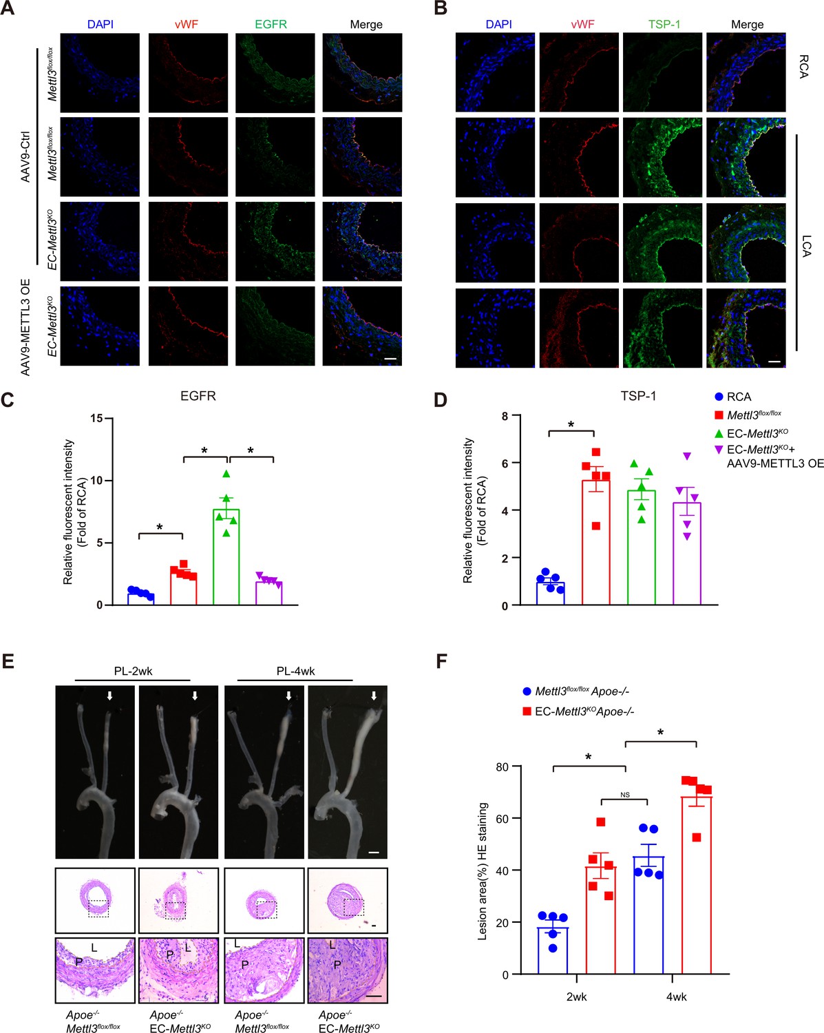
Epidermal growth factor receptor (EGFR) contributes to EC activation in endothelial methyltransferase like 3 (Mettl3)-deficient mice.

(A–B) EC-Mettl3KO and Mettl3flox/flox mice underwent partial ligation of the carotid artery for 2 weeks were infused with the indicated adeno-associated virus. Immunofluorescence staining for expression of EGFR, thrombospondin-1 (TSP-1) in ECs of the carotid artery of mice. Scale bar, 80 μm. (C–D) Quantification of the relative fluorescence intensity of EGFR and TSP-1. Data are shown as the mean ± SEM, *p<0.05 (two-way ANOVA with Bonferroni multiple comparison post hoc test). n = 5 mice. (E) Eight-week-old male Apoe–/–Mettl3flox/flox and Apoe–/– EC-Mettl3KO mice with 2 or 4 weeks of partial ligation were fed a Western-type diet, and arterial tissues were isolated to examine atherosclerotic lesions. Scale bar: 1.5 mm. Ligated coronary arteries were sectioned for hematoxylin-eosin staining. Scale bar: 100 μm. L, lumen; P, plaque. (F) Quantification of lesion area. Data are shown as the mean ± SEM, *p<0.05, NS, not significant (one-way ANOVA with Bonferroni multiple comparison post hoc test). n = 5 mice.

Figure 5—source data 1

EGFR contributes to EC activation in endothelial Mettl3-deficient mice.

https://cdn.elifesciences.org/articles/69906/elife-69906-fig5-data1-v2.zip

Mettl3-deficient ECs accelerates atherosclerosis in Apoe-/- mice

To further detect the proatherogenic role of endothelial METTL3, we carried out a standard atherosclerosis study in Apoe-/- EC-Mettl3KO mice and Apoe-/- Mettl3flox/flox mice. After 12 weeks of WTD, Oil Red O staining of aortas revealed that, in comparison with Apoe-/- Mettl3flox/flox mice, Mettl3-deficient ECs significantly increased the total and AA atherosclerotic area in aortas (Figure 6A and B). Aortic root staining showed that Mettl3 deficiency increased the lesion area, lipid deposition, and macrophage infiltration, as well as EGFR and VCAM-1 expression, but had minimal effects on collagen fiber or vascular smooth muscle cell content (Figure 6C–H). These results indicate that METTL3 is an important effector in EC activation and atherogenesis.

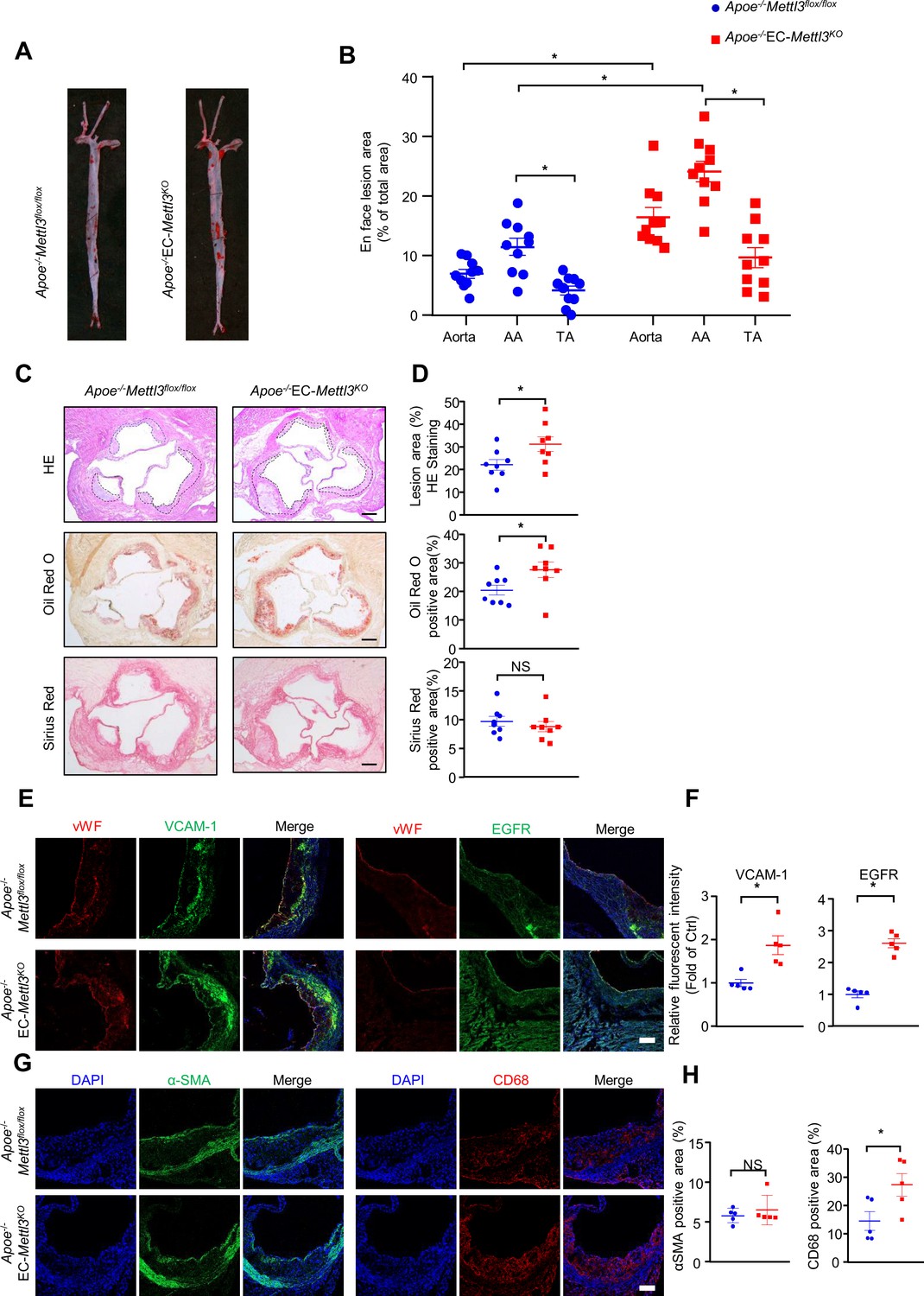
EC-specific methyltransferase like 3 (METTL3) deficiency accelerates atherogenesis in Apoe-/- mice.

Apoe-/-EC-Mettl3KO and Apoe-/-Mettl3flox/flox mice were fed a Western-type diet for 12 weeks. (A) Oil Red O staining of aortas. (B) Plaque area as a percentage of total area. AA, aortic arch; TA, thoracic aorta. Data are shown as the mean ± SEM, *p<0.05 (two-way ANOVA with Bonferroni multiple comparison post hoc test). n = 10. (C–D) HE, Oil Red O, and Sirius Red immunofluorescence staining of aortic roots. White dashed line indicates the size of plaque. Quantification of plaque size, Oil Red O-positive area in plaque size, Oil Red O-positive area in plaque and collagen fiber (Sirius Red). Data are shown as the mean ± SEM, *p<0.05 (Student’s t test). n = 8. (E and G) Epidermal growth factor receptor (EGFR), vascular adhesion molecule 1 (VCAM-1), vWF, α-SMA, and CD68 immunofluorescence staining of aortic roots. Scale bar, 20 μm. (F and H) Quantification of the relative fluorescence intensity of VCAM-1, EGFR, α-SMA, and CD68. Data are shown as the mean ± SEM, *p<0.05, NS, not significant (Student’s t test). n = 5.

Figure 6—source data 1

EC-specific METTL3 deficiency accelerates atherogenesis in Apoe-/- mice.

https://cdn.elifesciences.org/articles/69906/elife-69906-fig6-data1-v2.zip

TSP1/EGFR signaling is involved in atherosclerosis

To investigate the function of the endothelial TSP1/EGFR axis in atherogenesis, the endothelium of Apoe-/- EC-Mettl3KO and Apoe-/- Mettl3flox/flox mice with a partially ligated carotid artery was infected with lentivirus-mediated Thbs1 shRNA. The knockdown efficiency of Thbs1 shRNA was confirmed by en face staining (Figure 7—figure supplement 1A-B). Two weeks after ligation, the lesion area in Apoe-/- EC-Mettl3KO mice was significantly increased by 2.5-fold compared to that in Apoe-/- Mettl3flox/flox mice. However, Thbs1 knockdown successfully reversed the lesion area in both genotypes of mice (Figure 7A and B). The EGFR-selective tyrphostin AG1478 significantly reduced the phosphorylation of EGFR without affecting the total EGFR levels in both genotypes of mice (Figure 7—figure supplement 1C-E). Consistent with Thbs1 knockdown, AG1478 exerted the same anti-atherosclerotic effect (Figure 7A and B). Both of them inhibited the expression of VCAM-1 induced by EC Mettl3 deficiency in partially ligated carotid artery (Figure 7C and D). The levels of plasma triglyceride and cholesterol were unchanged among the groups (Figure 7—figure supplement 1F). These data indicate that endothelial Mettl3-mediated m6A in atheroprone regions is dependent upon TSP-1/EGFR in vivo.

Figure 7 with 1 supplement see all
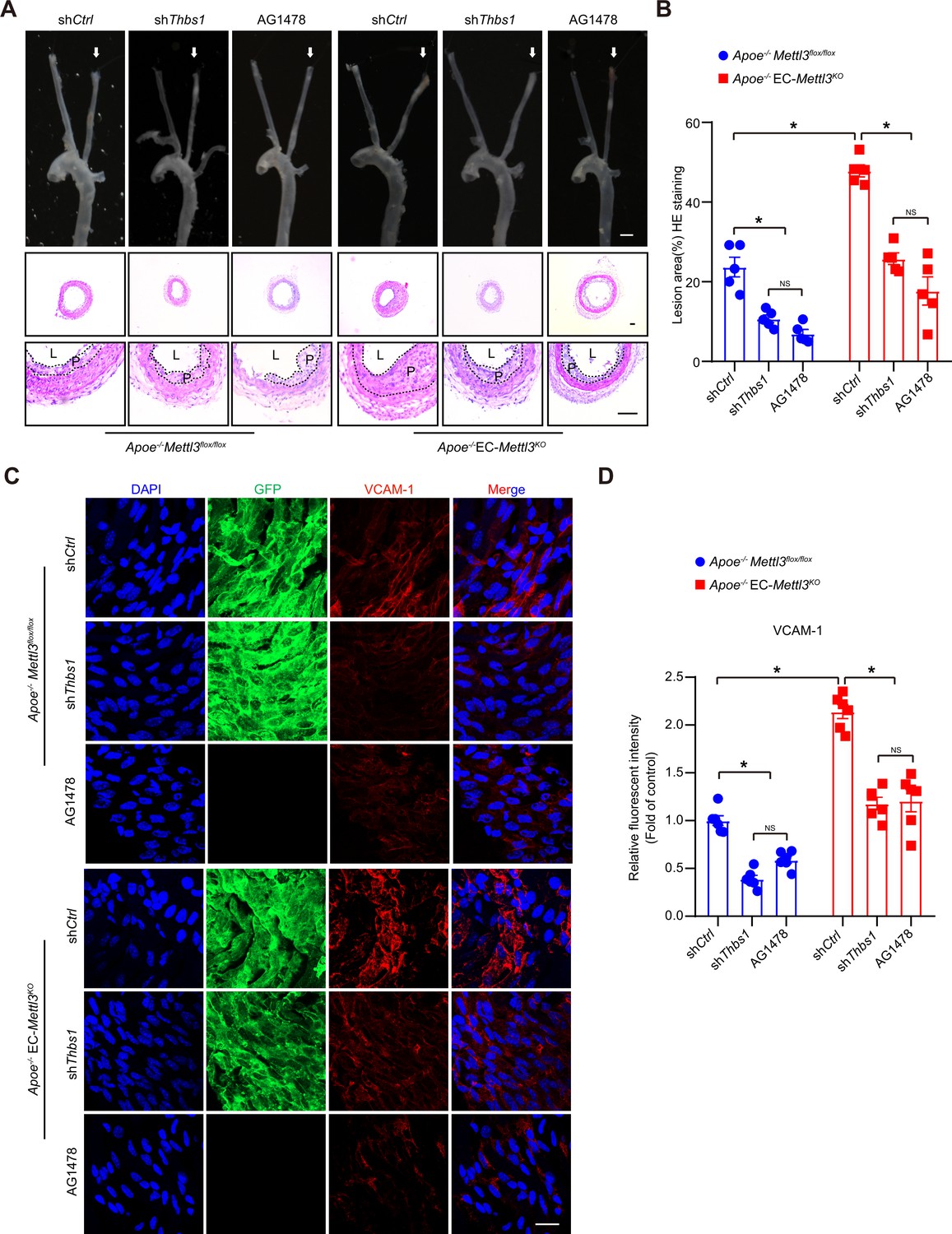
Thrombospondin-1/epidermal growth factor receptor (TSP1/EGFR) signaling is involved in atherosclerosis.

(A) An 8-week-old male Apoe–/– Mettl3flox/flox and Apoe–/– EC-Mettl3KO mice with 2 weeks of partial ligation were infused with the indicated lentiviruses or pretreated with AG1478 (AG, 10 mg/kg/day) for 7 days, and arterial tissues were isolated to examine atherosclerotic lesions. Scale bar: 1.5 mm. Ligated coronary arteries (LCAs) were sectioned for hematoxylin-eosin (H&E) staining. Scale bar: 100 μm. L, lumen; P, plaque. (B) Quantification of lesion area. Data are shown as the mean ± SEM, *p<0.05, NS, not significant (two-way ANOVA with Bonferroni multiple comparison post hoc test). n = 5 mice. (C) Apoe–/– Mettl3flox/flox and Apoe–/– EC-Mettl3KO mice with 2 weeks of partial ligation were infused with the indicated lentiviruses or pretreated with AG1478 (AG, 10 mg/kg/day) for 7 days. En face immunofluorescence staining of the expression of vascular adhesion molecule (VCAM-1) in ECs of the carotid artery of mice. Scale bar, 20 μm. (D) Quantification of the relative fluorescence intensity of VCAM-1. Data are shown as the mean ± SEM, *p<0.05, NS, not significant (two-way ANOVA with Bonferroni multiple comparison post hoc test). n = 6.

Figure 7—source data 1

TSP1/EGFR signaling is involved in atherosclerosis.

https://cdn.elifesciences.org/articles/69906/elife-69906-fig7-data1-v2.zip

Discussion

Our results reveal that RNA m6A modification plays an important role in regulating endothelial activation and the atherogenesis response to oscillatory flow. First, we found that m6A modification levels were reduced concomitant with downregulation of METTL3 in atheroprone regions. Second, oscillatory flow-derived MeRIP-seq analysis revealed that m6A modification of the EGFR 3’UTR contributes to atherogenesis. Third, overexpression of METTL3 restored m6A modification and reversed VCAM-1 expression and the monocyte adhesion ability response to OS. Finally, TSP-1/EGFR inhibition prevented the development of atherosclerosis, suggesting a novel therapeutic method for atherosclerosis patients. Together, our findings demonstrate that METTL3 and m6A modifications could alleviate endothelial activation and atherogenesis through accelerated degradation of oscillatory flow-induced EGFR mRNA expression.

Atherosclerotic plaques tend to develop in the vasculature locations with increased shear stress and OS. EC activation in response to oscillatory flow plays important roles in regulating circulatory functions and atherosclerosis development, which is involved in the expression of various genes (Li et al., 2019; Zhou et al., 2014). Recently, increasing evidence has suggested that m6A mRNA modification participates in a number of biological functions and in progression of ECs (Shi et al., 2019; Wang et al., 2014; Xiang et al., 2017). In this study, we generated tamoxifen-inducible endothelium-specific Mettl3 deficiency mice with or without Apoe-/- background. Western blot analysis of arterial intima revealed decreased METTL3 in AA compared to TA in mice of both genotypes (Figure 2A). Overexpression of Mettl3 has potential protective effects on endothelial activation in atheroprone regions (Figure 2F), indicating the involvement of both m6A modification and METTL3 in atherogenesis.

As the core methyltransferase subunit, METTL3 has been reported to modulate embryonic development (Aguilo et al., 2015) and spermatogenesis (Xu et al., 2017), and its deletion in mice causes early embryonic lethality (Geula et al., 2015). METTL3 also plays a central role in osteogenic differentiation and inflammatory response (Wu et al., 2018; Zhang et al., 2019), which act similarly in ECs. METTL3 expression is upregulated during osteoblast differentiation and downregulated after LPS (lipopolysaccharide) stimulation, while its depletion enhances the expression of Smad genes and proinflammatory cytokine expression in MAPK and NF-κB signaling pathways (Zhang et al., 2019). Chien et al. proposed that METTL3 is increased after long term of disturbed flow for 48 hr (Chien et al., 2021). We performed a short term of disturbed flow for 6 hr, and the mechanism research in genotype mice limited in 2 or 4 weeks. Furthermore, most evidence was demonstrated in EC-specific Mettl3 knockout mice. We presume a possibility that the differential expression of METTL3 is attributed to variations in the stimulus, such as treatment time and strength. The development of hematopoietic stem cells (HSPCs) also requires METTL3-mediated m6A modification, and mettl3 deficiency in arterial endothelial cells blocks endothelial-to-hematopoietic transition, followed by suppression of the generation of HSPCs through activation of Notch signaling (Zhang et al., 2017). METTL3 plays a similar role in cerebral arteriovenous malformation, and deletion of METTL3 in ECs significantly affects angiogenesis by reducing heterodimeric Notch E3 ubiquitin ligase formed by DTX1 and DTX3L (Wang et al., 2020). METTL3 seems to be essential for maintaining endothelial function. Here, we analyzed RNA-seq and MeRIP-seq to reveal declined methylation and increased expression of cell adhesion- and migration-related pathways. Our results describe the role of m6A and METTL3 in atherogenic progression and identify therapeutic strategies against atherosclerosis through m6A modification and its related targets.

It has been reported that EGFR plays a role in foam cell transformation and accelerates atherosclerotic lesions characterized by accumulation of smooth muscle cells and macrophages (Wang et al., 2017). Although EGFR is expressed at low levels in ECs, explosive elevation of transcription and phosphorylation of EGFR were detected after OS treatment (Figure 3D and Figure 4), consistent with a report from Rizvi et al., 2013. METTL3 binds to EGFR mRNA near the stop codon and is responsible for its m6A modification, modulating EGFR mRNA stability in human cancer cells (Lin et al., 2016). Chien et al. also pointed out EGFR as a hypomethylated gene in METTL3 knockdown under OS (Chien et al., 2021). Our results demonstrated that the EGFR 3’UTR response to OS and double mutation of the 3’UTR abolished the reduction induced by METTL3 overexpression in ECs (Figure 3), indicating that EGFR is responsible for m6A modification in response to OS treatment. EGFR is activated in response to its EGF-like repeats, such as TSP1, which require matrix metalloprotease activity, and the ligand-binding portion of the EGFR ectodomain (Liu et al., 2009). The EGFR signaling pathway was blocked by overexpression of METTL3 in response to OS and supplementation with TSP-1 (Figure 4A–D). However, TSP-1 was not affected by METTL3 intervention, and we presume that TSP-1 acts as a specific ligand that activates EGFR after OS treatment. Moreover, the EGFR selective tyrphostin AG1478 inhibited EGFR phosphorylation at tyrosine 1068 without affecting the total levels of EGFR (Garg et al., 2011; Wang et al., 2017) and had an atheroprotective role (Wang et al., 2017). AG1478 reduced levels of p-EGFR and downstream proteins in the presence of OS, and Thbs1 shRNA and AG1478 reversed atherogenesis in partially ligated arteries in EC-Mettl3KO mice (Figure 4 and Figure 7).

In summary, we provide both in vitro and in vivo evidence demonstrating that m6A RNA modification regulates progression of EC activation in response to the onset of OS and subsequently, early atherogenesis. It should be noted that METTL3-modified EGFR mRNA participates in the pathogenic mechanism of atherogenesis. The TSP-1/EGFR axis may contribute to m6A-modified atherogenesis, and inhibition of the axis helps to retard atherosclerosis.

Materials and methods

Key resources table
Reagent type (species) or resourceDesignationSource or referenceIdentifiersAdditional information
Antibodyanti-METTL3(Rabbit monoclonal)Cell Signaling TechnologyCat# 96,391WB (1:1000)
Antibodyanti-METTL14(Rabbit monoclonal)Cell Signaling TechnologyCat# 51,104WB (1:1000)
Antibodyanti-METTL16(Rabbit monoclonal)Cell Signaling TechnologyCat# 17,676WB (1:1000)
Antibodyanti-WTAP(Rabbit monoclonal)Cell Signaling TechnologyCat# 56,501WB (1:1000)
Antibodyanti-Virillizer(Rabbit monoclonal)Cell Signaling TechnologyCat# 88,358WB (1:1000)
Antibodyanti-phospho-EGFR(Rabbit monoclonal)Cell Signaling TechnologyCat# 3,777WB (1:1000)
Antibodyanti-EGFR(Rabbit monoclonal)Cell Signaling TechnologyCat# 4,267WB (1:1000)
Antibodyanti-VCAM-1(Rabbit monoclonal)Cell Signaling TechnologyCat# 15,631WB (1:1000)
Antibodyanti-VCAM-1(Rabbit monoclonal)Cell Signaling TechnologyCat# 39,036IF (1:100)
Antibodyanti-thrombospondin-1(Rabbit monoclonal)Cell Signaling TechnologyCat# 37,879WB (1:1000)
Antibodyanti-αSMA(Rabbit monoclonal)Cell Signaling TechnologyCat# 19,245WB (1:1000)
Antibodyanti-phospho-ERK(Rabbit monoclonal)Cell Signaling TechnologyCat# 8,544WB (1:1000)
Antibodyanti-phospho-AKT(Rabbit monoclonal)Cell Signaling TechnologyCat# 5,012WB (1:1000)
Antibodyanti-ERK(Mouse monoclonal)Santa CruzCat# sc-271269WB (1:1000)
Antibodyanti-AKT(Mouse monoclonal)Santa CruzCat# sc-5298WB (1:1000)
Antibodyanti- METTL3(Rabbit monoclonal)ProteintechCat# 15073–1-APIF (1:100)
Antibodyanti- GFP(Rabbit monoclonal)ProteintechCat# 50430–2-APIF (1:100)
Antibodyanti- GAPDH (Rabbit monoclonal)ProteintechCat# 60004–1-IgWB (1:5000)
Antibodyanti- EGFR (Rabbit monoclonal)AbcamCat# ab52894IF (1:100)
Antibodyanti- VE-cadherin (Rat monoclonal)AbcamCat# ab33168IF (1:100)
Antibodyanti- CD31(Rabbit monoclonal)AbcamCat# ab24590IF (1:100)
Antibodyanti- CD68(Rabbit monoclonal)AbcamCat# ab955IF (1:100)
Antibodyanti- vWF(Sheep monoclonal)AbcamCat# ab11713IF (1:100)
Antibodyanti-thrombospondin-1(Mouse monoclonal)AbcamCat# ab1823IF (1:100)
Antibodyanti- thrombospondin-1 (Mouse monoclonal)AbcamCat# ab1823IF (1:100)
AntibodyAlex 488-conjugated goat anti-rabbit antibodyThermo Fisher ScientificCat# A-11008IF (1:200)
AntibodyAlex 594-conjugated goat anti-mouse antibodyThermo Fisher ScientificCat# A-11008IF (1:200)
AntibodyAlex 488-conjugated goat anti-rabbit antibodyThermo Fisher ScientificCat# A-11005IF (1:200)
AntibodyAlex 594-conjugated donkey anti-sheep antibodyThermo Fisher ScientificCat# A-11016IF (1:200)
Chemical compound, drugAG1478SelleckCat# S2728
Chemical compound, drugRecombinant Human Thrombospondin-1AbsinCat# abs 046651.03 mg/ml
sequence-based reagentHuman EGFR-3utrThis paperN/ASequences in Supplementary file 3
sequence-based reagentHuman EGFRThis paperN/ASequences in Supplementary file 3
sequence-based reagentHuman THBS1This paperN/ASequences in Supplementary file 3
sequence-based reagentHuman GAPDHThis paperN/ASequences in Supplementary file 3
software, algorithmIngenuity Pathway AnalysisNational Clinical Research Center for Blood Diseaseshttp://www.ingenuity.com/
software, algorithmPrism version 8.0GraphPadSoftware Inchttps://www.graphpad.com/scientific-software/prism/

Cell Culture and Shear Stress Experiments

Request a detailed protocol

HUVECs were isolated and cultured as described (He et al., 2018; Li et al., 2019). Mouse aortic endothelial cells (catalog no. CP-M075) and specific medium (CM-M075) were purchased from Procell (Wuhan, China). For flow experiments, confluent monolayers of HUVECs were seeded on glass slides, and a parallel plate flow system was used to impose oscillatory flow (0.5 ± 4 dyn/cm2). THP-1 cells (ATCC, catalog TIB-202) were cultured with 1640 medium supplemented with 10% fetal bovine serum. The flow system was enclosed in a chamber held at 37°C and ventilated with 95% humidified air plus 5% CO2.

Recombinant Mettl3 adenovirus, adeno-associated virus, and THBS1 lentivirus construction

Request a detailed protocol

Adenoviruses expressing green fluorescent protein and Ad-Flag-tagged human METTL3 (Ad-METTL3) were purchased from GeneChem (Shanghai, China). Recombinant AAV serotype nine vectors carrying METTL3 or empty vector were manufactured by GeneChem Co, Ltd (Shanghai, China). Lentiviruses carrying short hairpin RNA (shRNA) targeting Thbs1 (LV-shThbs1) and nonspecific shRNA (LV-shCtrl) were constructed by Shanghai Genechem Co (Shanghai). HUVECs were infected with adenovirus at a multiplicity of infection (MOI) of 10, and no detectable cellular toxicity was observed.

Western blot analysis

Request a detailed protocol

Cells or tissues were homogenized in cold RIPA lysis buffer supplemented with complete protease inhibitor cocktail and phosSTOP phosphatase inhibitor (Roche). Proteins were resolved by SDS-PAGE and transferred to NC membranes (Bio-Rad). Target proteins were detected using specific primary antibodies (1:1000). Bound antibodies were detected by horseradish peroxidase-conjugated secondary antibody (1:5000) and visualized by enhanced chemiluminescence (Cell Signaling Technology).

Cell adhesion assay

View detailed protocol

THP-1 cells were labeled with CellTrace calcein red-orange AM (Thermo Fisher catalog C34851) and then plated onto HUVEC plates at 2 × 10⁶ cells/well. After incubation for 60 min at 37°C, nonadherent cells were removed by three washes with phosphate buffered saline (PBS). The numbers of stained adherent cells in five random fields were counted for each group under a fluorescence microscope.

Immunofluorescence staining

Request a detailed protocol

Arteries or aorta sections were fixed with 4% paraformaldehyde for 15 min. After permeabilization/blocking in 0.05% Triton X-100 (in PBS) and 1% bovine serum albumin (BSA) for 30 min at room temperature, aortas were incubated at 4°C overnight in incubation buffer containing 1% BSA and primary antibodies (1:100) against METTL3, p-EGFR, EGFR, VCAM-1, GFP, αSMA, CD68, vWF, TSP-1, and CD31. After washing in PBS three times, aortas were incubated with Alexa Fluor 488- or Alexa Fluor 594-conjugated secondary antibodies (1:200) for 1 hr at room temperature. Fluorescent signals were detected by using a Zeiss confocal laser scanning microscope.

Animals

We established tamoxifen-inducible EC-specific Mettl3-deficient (EC-Mettl3KO) and littermate control (Mettl3flox/flox) mice. Mice carrying the floxed Mettl3 allele mice were crossed with mice harboring Cre recombinase under the control of the Cdh5 promoter, which contained a tamoxifen-inducible EC-specific Cre. Tamoxifen was administered once every 24 hr for five consecutive days. All mice were on a C57BL/6 J background and were maintained under a 12:12 hr light/dark cycle (lights on at 7:00 and lights off at 19:00). The investigation conformed to the Guide for the Care and Use of Laboratory Animals by the US National Institutes of Health (NIH Publication No. 85–23, revised in 2011). All study protocols and the use of animals were approved by the Institutional Animal Care and Use Committee of Tianjin Medical University.

Animal experiments

Request a detailed protocol

Partial ligation of carotid artery was performed as described (Zhang et al., 2020). The indicated mice were anesthetized by using isoflurane (2–3%). Carotid arteries of both sides were exposed by creating a ventral midline incision (4–5 mm) in the neck. The left external carotid, internal carotid, and occipital arteries were ligated and right external carotid as sham; the superior thyroid artery was left intact. Mice were monitored until recovery in a chamber on a heating pad after surgery. Mice were fed WTD for 2 and 4 weeks. For lentivirus infection studies, a single exposure of 5 × 10⁵ TU adenovirus was lumenally delivered to the left carotid artery and kept inside for 40 min to allow for sufficient infection. The lentivirus solution was subsequently removed and blood flow was restored. For AAV9 infection studies, AAV9-TIEp-METTL3/empty vectors (3 × 10¹¹ vector genomes/mice) were delivered by intravenous injection. One week after AAV9 delivery, partial ligation was performed. Mice were fed WTD immediately after the surgery for 2 weeks.

Quantification of lipid levels

Request a detailed protocol

Blood samples were collected by tail bleeding into heparin-coated tubes. Plasma was separated by centrifugation. Total plasma cholesterol, triglycerides, low-density lipoprotein-cholesterol, and high-density lipoprotein-cholesterol levels were measured by using kits from BioSino Bio-Technology and Science Inc (Beijing, China).

Total RNA extraction and real-time quantitative PCR analysis

Request a detailed protocol

Total RNA was extracted from tissue and HUVECs using RNA extraction kits (TransGen Biotech, ER501-01, China). Complementary DNA was synthesized using reverse transcription using SuperScript III and random primers (Thermo Fisher, catalog 12574035, MA). Real-time qPCR was performed using the Brilliant II SYBR Green qPCR Master Mix (Stratagene, CA) and the ABI 7900HT Real-Time PCR System (Life Technologies, CA). The results were normalized to those of 18 S. Data were calculated using comparative Ct values. The primer sequences are listed in Supplementary file 3.

RNA purification and mRNA purification

Request a detailed protocol

Total RNA was extracted using TRIzol (Thermo Fisher), treated with Turbo DNase (Thermo Fisher), and then subjected to mRNA purification with the Dynabeads protein A mRNA purification kit (Thermo Fisher) following the manufacturer’s instructions.

UHPLC-MRM-MS/MS analysis of mononucleotides

Request a detailed protocol

An amount of 200 ng purified mRNA of each sample was digested with 0.1 U Nuclease P1 (Sigma, catalog N863) and 1.0 U calf intestinal phosphatase (New England Biolabs, catalog M0290), in the final reaction volume of 50 μl and incubated at 37°C for over 5 hr. The mixture was then filtered with ultra-filtration tubes (MW cutoff: 3 KDa, Pall, Port Washington, NY) through centrifuging at 14,000 g for 20 min. The samples were subjected to UHPLC-MRM-MS/MS analysis for detection of m6A and rA. The UHPLC-MRM-MS/MS analysis was performed according to the previous report (Xiao et al., 2016).

RNA-seq

Request a detailed protocol

RNA-seq libraries were directly constructed using the KAPA RNA HyperPrep Kit (KAPA Biosystems) following the manufacturer’s instructions. Sequencing was performed on an MGISEQ-2000 platform with a single end 50 nt read length. Three replicates were used in RNA-seq studies.

MeRIP-seq

Request a detailed protocol

MeRIP was performed according to previously described methods. First, 5 μg of anti-m6A antibody (Synaptic Systems, 202003) was incubated with 20 μL Dynabeads Protein A (Invitrogen, 1,001D) in 500 μL IPP (immunoprecipitation buffer) buffer (150 mM NaCl, 10 mM Tris-HCl, pH 7.4, 0.1% NP-40, 10 U RNase Inhibitor) at 4°C for 1 hr. Second, ~ 100 ng of fragmented mRNA was added to the antibody-bead mixture and incubated at 4°C for 4 hr with gentle rotation. After extensive washing with IPP buffer, high-salt wash buffer (500 mM NaCl, 10 mM Tris-HCl, pH 7.4, 0.1% NP-40), and low-salt wash buffer (50 mM NaCl, 10 mM Tris-HCl, pH 7.4, 0.1% NP-40), RNA fragments were eluted from the beads with proteinase K (Roche, 3115836001) digestion at 55°C for 1 hr and extracted with phenol-chloroform extraction and ethanol precipitation. The recovered RNAs were subjected to library preparation using the KAPA RNA HyperPrep kit (KAPA Biosystems, KK8541). Sequencing was performed on an Illumina HiSeq X-Ten platform with paired end 150 base pair (bp) read length.

Sequencing data analysis

Request a detailed protocol

For general preprocessing, SOAPnuke (version 1.5.2) and Trim Galore (version 0.6.4) were used to trim off the adapter sequences and low-quality bases for all samples. The remaining reads were aligned to the human genome (version hg19) using Hisat2 (version 2.0.5) (Kim et al., 2015), and only uniquely mapped reads with q ≥ 20 were used in subsequent analysis.

For MeRIP-seq, two biological replicates were conducted. The replicates of each sample were merged for m6A peak calling using MACS2 (version 2.1.4) (Zhang et al., 2008) with the corresponding input samples as a control. Default parameters were used, except for ‘-nomodel –keep dup all’, to turn off fragment size estimation and to keep all uniquely mapped reads in MACS2. Finally, each peak was annotated based on Ensembl (release 72) gene annotation information using BETools intersectBed (version 2.28.0) (Quinlan and Hall, 2010). For RNA-seq, the number of reads mapped to each gene (Ensembl 72) was counted using featureCounts (version 1.6.2) (Liao et al., 2014) with the default parameters, except for ‘-s 2’.

Statistical analysis of differentially expressed genes and gene ontology analysis

Request a detailed protocol

Differentially expressed genes between ST and OS treatments were identified using DEGseq (Wang et al., 2010). Differentially expressed genes were identified by log2 |fold change| > log2 |1.5| and FDR < 0.05. GO analysis was performed using DAVID (https://david.ncifcrf.gov/), and p < 0.05 were considered significantly enriched.

Motif identification within m6A peaks and differential m6A peaks

Request a detailed protocol

HOMER (version 4.7) (Heinz et al., 2010) was used to identify the motif enriched by the m6A peak, and the motif length was limited to seven nt. Peaks annotated to mRNA were considered target sequences, and background sequences were constructed by randomly perturbing these peaks using shuffleBed of BEDTools (version 2.28.0) (Quinlan and Hall, 2010). Based on the enrichment level, differential m6A peaks were identified as those with log2 |fold change| > log2 |1.5|.

Statistics

Sample sizes were designed with adequate power according to the literature and our previous studies. No sample outliers were excluded. Experiments were not randomized, and the investigators were not blinded to allocation during experiments or outcome assessment. The variance between the groups being statistically compared was similar. Data are presented as mean ± SEM. Statistical analysis was performed using GraphPad Prism 8. All the data with n ≥ 6 was tested for normality using the Shapiro-Wilk normality test. For normally distributed data, comparisons between two groups were performed using unpaired Student’s t test, and comparisons among three or more groups were performed using one-way or two-way ANOVA followed by Bonferroni’s multiple comparisons correction; For non-normally distributed data and the data with n < 6, Mann-Whitney U test or the Kruskal-Wallis test followed by Dunn’s multiple comparison tests were performed as appropriate. For the immunofluorescence images, quantification was normalized as interest of district per area. Three to five images per aorta and region of interest of each image were used for analysis. In all experiments, p<0.05 was considered statistically significant.

Data availability

RNA-seq and MeRIP-seq data generated in this study have been deposited to the Genome Sequence Archive in BIG Data Center under accession number PRJCA004746.

The following data sets were generated
    1. Liang D
    (2022) Genome Sequence Archive
    ID PRJCA004746. RNA N6-methyladenosine modulates endothelial atherogenic responses to disturbed flow.

References

Decision letter

  1. Edward A Fisher
    Reviewing Editor; New York University Grossman School of Medicine, United States
  2. Mone Zaidi
    Senior Editor; Icahn School of Medicine at Mount Sinai, United States

In the interests of transparency, eLife publishes the most substantive revision requests and the accompanying author responses.

Decision letter after peer review:

Thank you for submitting your article "RNA N6-methyladenosine modulates endothelial atherogenic responses to disturbed flow" for consideration by eLife. Your article has been reviewed by 3 peer reviewers, one of whom is a member of our Board of Reviewing Editors, and the evaluation has been overseen by Mone Zaidi as the Senior Editor. The reviewers have opted to remain anonymous.

The reviewers have discussed their reviews with one another, and the Reviewing Editor has drafted this to help you prepare a revised submission.

Essential revisions:

You will see in the reviewers' comments numerous questions and suggestions. After consultation among the reviewers, we have listed the essential items that need to be addressed in a revised manuscript:

1) The evidence that mettl3 is downregulated with shear stress is not convincing. The western blot in Figure1a shows very subtle differences in mettl3 and many would question how the change proposed here modulates physiologic changes in m6A levels. To be convincing the authors should measure m6A levels as noted in Reviewer 2, point #1. This can be done using a commercial kit or preferably mass spec which is widely available from many groups now. Reviewer 3 also was concerned about m6A levels, and noted that the lack of m6A changes in vivo needs to be resolved, since altered m6A levels are the proposed mechanism of action.

2) The wild animals have almost undetectable mettl3 levels so they are practically

"Knockouts". Then what is the basis for the huge differences in the plaque burden?

5) What is the justification that the authors have to explain that all of the effects observed in AKT, ERK and VCAM-1 under OS are corrected by EGFR inhibition? The data supporting this conclusion is lacking.

6) The discussion of the Chien et al. PNAS 2021 is very narrow, especially considering the major discrepancies between the studies. The authors only note that the difference in the in vitro studies may be from shorter vs. longer exposure to shear stress, but his cannot be the explanation for the discrepant results in vivo using similar models. Thus, a full comparison needs to be part of the Discussion.

7) Also, there are a number of technical issues, including the lack of controls, the choice of HUVECS vs. more relevant aortic ECs, as well as statistical issues the reviewers have raised that need to be addressed.

Reviewer #1:

There is a lot of interesting information in this manuscript. Most of the studies in vivo have looked at METTL3 deficiency, so I think that the protective role of sustaining its expression in the ligation model should be explored in addition. Also, given the many RNAs found to be modified by METTL3, I find it surprising that it mainly comes down to EGFR in ECs, and that a major player is TSP1. TSP1, for example, has been investigated in atherosclerosis for its effects in two other major cells types in plaques, namely SMC and macrophages. Attributing all of its effects to what it does to ECs may reflect the model they use, which some consider as an "advanced atherosclerosis" process (it certainly has disturbed flow), but given that they have apoE-/- mice, a more standard atherosclerosis study could have been performed. Another technical note is that HUVECs are controversial in their direct relevance to arterial ECs. A final note is that there is a competing paper earlier this year in which some of the important results are discrepant, so there is a need for reconciling the findings.

The authors have provided a lot of detailed and interesting data to support their contention that the methylation of the RNA encoding EGFR is a major regulator of a vascular pathology related to atherosclerosis. There are some issues and gaps, however, in the story, including:

1. A lot of the studies are done in HUVECs. Historically, this cell model has been used for EC studies, but it is well appreciated that the direct relevance to arterial ECs is not clear. Were studies in other cell models of ECs performed, with similar results for key findings replicated?

2. TSP-1 has been studied for its effects on macrophages and SMCs, with reports to support its effects on atherosclerosis working through these cell types. The authors imply that all of the effects in their system is through the EC EGFR pathway. Is this perhaps a reflection of the ligation model they use? It has many notable differences from a standard model of atherosclerosis, so are the effects that seem to be confined to ECs model specific? With having apoE-/- mice available, I am surprised a standard atherosclerosis study was not performed.

3. The overexpression of METTL3 in vitro appeared to be protective against OS. While the authors provide experimental data in vivo with siRNA to further knock it down, I would think that an in vivo protection study would be a valuable addition. Also, with regard to the lentivirus experiment, how was it excluded that effects on other cell types (besides ECs) were not contributory?

4. THP-1 adhesion studies were done in some places, but the early studies referring to EC activation were based on the effects on adhesion molecules (e.g., VCAM). Some functional studies should have been done to show activation had a consequence on monocyte adhesion.

5. What is the basis for the effects of METTL3 changes on EGFR phosphorylation and the other signaling molecules reported on?

6. Going back to the specificity issue, there were many changes in the "methylome"- it is surprising that the major impact is exclusive to EGFR.

7. How do the authors explain the differences with the findings in Chien et al. (PNAS 118:e2025070118, 2021)?

Reviewer #2:

A notable strength of the current study is leveraging unbiased m6A interrogation approaches to decipher mechanisms of shear stress induced endothelial programming which is of substantial interest. Although others reported on generation of endothelial-specific Mettl3 KO, this is not a trivial endeavor and adds to the significance. It is difficult to ignore however that the most important conclusions of the paper were reported in a highly similar recent manuscript (Chien et al. PNAS. 2021). Notably the study from Shu Chien's group performed a rigorous interrogation of endothelial responses in response to oscillatory flow using eCLIP to map m6A sites, identified the reader protein involved and used a similar in vivo atherogenesis model perturbing Mettl3 to validate their findings. Implicating the EGFR signaling pathways and use of rescue/epistatic studies here is a novel aspect but the overall conceptual advance may be somewhat incremental.

1. The authors claim that mettl3 levels are reduced in response to OS but they provide no evidence that the subtle changes in mettl3 in endothelial cells(see figure 1A) translates in to meaningful changes in m6A levels. Thus, a major limitation of the work is that there are no direct m6A measurements? The authors need to measure global m6A levels preferably by mass spectrometry under different conditions. Measurement of m6A levels should also be done in the mettl3 overexpression studies.

2. Related to point#1, the proposed regulation of mettl3 in endothelial is opposite to previous work which showed that mettl3 is not downregulated and in fact upregulated in response to OS and other pro-atherogenic conditions (PMID: 33579825 and 32755566). The authors claim that these findings may be due to differences in conditions but this not well explored. The authors need to better consolidate their findings with previous work and ideally show experimentally the basis for Mettl3 regulation.

3. There are important details missing for key studies. How many replicates were used for the m6A-seq studies? How are the peaks shown in Figure 3C normalized? Can the authors show additional m6A peaks in the supplement? Can the authors also show a more expanded list of enriched motifs under different conditions and not just the top one?

4. Similar to the question above can the authors provide more details on number of replicates for RNA-seq study?

5. For some panels there appears to be dramatic differences in Mettl3 levels in the in vivo model. For example, in IHC staining shows that Mettl3 is almost completely absent in the LCA model (Figure 2D, and Supp Figure 5A). How do authors explain the big differences in plaque burden in Figure 5 if control animals are practically Mettl3 deficient? Curiously staining for Mettl3 is absent in figure 5.

6. The differences in motifs show in Figure 3B is an interesting finding but the physiologic significance of this finding is unclear since it invokes that the "interactome" of methyltransferase complex may be different depending on conditions which is not proven. Suggest minimizing this point in the text since its seems distracting.

7. Ref cited twice p28 line 7 and 10.

8. Please provide legends for abbreviations in figures.

Reviewer #3:

In the present work, Bochuan Li et al. studied the role of endothelial RNA N6-methyladenosine in atherogenesis. Using in vitro and in vivo approaches, Li and collaborators have shown that disturbed flow decreases the expression of methyltransferase METTL3 in endothelial cells. METTL3 is the enzyme responsible for nearly all the N6-methyladenosin (m6A) addition in mRNAs. Using in vitro methods, the authors show that under conditions of shear stress, human umbilical vein endothelial cells (HUVECs) demonstrate reduced m6A modification in the EGFR mRNA, which they associate with reduced EGFR mRNA degradation. Additionally, the authors used partial ligation of the left common carotid artery, which is an animal model of disturbed flow, to show that METTL3 expression is decreased with increased EGFR levels in the endothelium. They also demonstrate that the increase in EGFR expression promotes vascular inflammation and atherosclerosis by upregulating vascular adhesion molecule 1 (VCAM-1). These findings were also observed using a METTL3 endothelial cell specific knockout animal model. Furthermore, both overexpression of METTL3, or pharmacological inhibition of EGFR signaling, reduced VCAM-1 expression. Based on these results, the authors conclude that vascular inflammation in areas with disturbed flow is regulated by mRNA m6A modification, and that METTL3-mediated EGFR mRNA modification participates in the pathogenic mechanism of atherogenesis.

The major strength of this study is that the authors used a variety of in vitro and in vivo models, combining genetic approaches (METTL3 specific KO and the overexpression of METTL3), and pharmacological approaches, to show that there is a role for METTL3 and EGFR in atherogenesis.

Despite many of the strengths that this work has, there are several key weaknesses that make their conclusions less convincing and will require additional experimentation to resolve. In particular, there are significant differences between the effect of perturbed flow on METLL3 expression observed in vitro and in vivo. While in vivo data showed a dramatic reduction of METTL3, the in vitro data showed a modest reduction in enzyme levels, and there was no difference in m6A modification between cells treated with normal or disturbed flow. The lack of m6A changes is most problematic and needs to be resolved, since altered m6A levels are the proposed mechanism of action. Fundamentally, it is not clear whether the in vitro perturbed flow model recapitulates the key properties of in vivo perturbed flow, or whether the in vivo effect is due to features other than altered flow.

Another factor that makes it difficult to interpret these data concerns the way that overexpression or silencing by lentiviral vector infection was carried out, including a lack of control studies. There are no data demonstrating that overexpression or silencing only occurs in the endothelium layer (which itself is doubtful). Therefore, it is difficult to interpret these results when it is possible that there are contributions from other cell compartments within the aortic wall, including vascular smooth muscle cells and recruited leukocytes. To more fully understand the results of this study, a more detailed description of the methods is needed, and a greater discussion of the study's findings is necessary. In addition, it is not clear how the quantification of fluorescence was done. Importantly, a proper statistical analysis is critical to evaluate these findings and determine whether certain conclusions are warranted. Another issue that needs to be corrected concerns the small number of animals used in each experiment. With the small number of animals used, it is not certain if the data follows a normal distribution prior to applying a parametric analysis (T-test or ANOVA). It is also not clear whether it is appropriate to to use the standard error of the mean (SEM) rather than standard deviation (SD) in many of these studies. SD seems to be proper analysis based on descriptions in experimental methods.

The work presented by Li et al. is meaningful and has a potential to be published, but at the present time it is incomplete and will require additional experimentation and clarification.

1. It will be more relevant for the study if the in vitro experiments are performed on aortic endothelial cells rather than HUVECs, especially since the authors are comparing and making correlations between in vivo and in vitro studies. in vivo studies are of course investigating aortic endothelial cells.

2. The lack of effect on m6A modification between cells treated with normal or disturbed flow is disconcerting, since that it is the activity of METTL3 that is proposed to be the mediator of the effects in this study. Consequently, assuming these data are correct, the authors should examine if there is an increase in METTL16 that could account for the fact that with lower METTL3 levels there are no changes in m6A levels. Another possible explanation could be that the remaining METTL3 enzyme is sufficient to carry out methyltransferase activity, or that loss of m6A requires considerably greater amount of time than considered in these studies. In that case, it would be important to add a METTL3 knockdown study as a control. A METTL3 knockdown would help to determine which changes in mRNA expression are due to METTL3 downregulation in OS conditions.

3. In Figure 3F, it is important to show the effect of METTL3 knockdown on EGFR mRNA stability. It is possible that the increase in the EGFR mRNA under OS could be a transcriptional effect. It needs to be determined whether under OS conditions, EGFR mRNA has a slower decay rate.

4. The authors showed that OS increases EGFR expression and signaling in HUVECs. Additionally, they showed that the activation of EGFR under OS increases AKT and ERK phosphorylation and an increase in VCAM-1 expression. Overexpression of METTL3 avoids the effects previously described and silencing of METLL3 recapitulates OS effects, and inhibition of EGFR phosphorylation with AG1478 impaired AKT and ERK phosphorylation and VCAM-1 expression under OS conditions. However, it is not clear why VCAM-1 protein levels increase under OS or with METTL3 silencing, and this needs to be resolved. Is it NF-κB or AP-1 mediated? What is the justification that the authors have to explain that all of the effects observed in AKT, ERK and VCAM-1 under OS are corrected by EGFR inhibition? The data supporting this conclusion is lacking. The authors seem to be suggesting that OS is sensed by HUVECs in an EGFR-mediated manner exclusively? That seems difficult to believe, and needs to be supported by additional evidence. Also, METTL3 overexpression and silencing must be demonstrated by immunoblots in Figure 4.

5. For the experiments with partial ligation of the left common carotid artery in which METTL3 endothelial cell specific knockout animals were used, the authors need to provide representative images of the immunofluorescence in the RCA of KO animals. This should also be quantified and compared statistically.

6. There is considerable background in a number of the images that makes it difficult to distinguish signal from noise, especially for CD31 (Figure 5). Because of this issue, it is difficult to conclude that VCAM-1 colocalizes with CD31 positive cells. Proper unambiguous staining is critical to demonstrate that the increase in VCAM-1 is in the endothelium and it is not also increasing in other cells like vascular smooth muscle cells that under inflammatory conditions express VCAM-1 as well. The best approach is to carry out staining in cross sections like the HandE staining studies. That way it will be clear if VCAM-1 expression is occurring exclusively in the endothelium.

7. For all studies in which overexpression or silencing was carried out, the staining should be shown in cross sections to demonstrate that the silencing or overexpression is limited to the endothelium and not to the intima or the media of the aortic wall. Was the silencing or overexpression homogenous along the whole endothelium?

8. Perturbed flow occurs naturally along the aorta, as the authors mentioned. One of these areas is the aortic root, in which atherosclerotic plaques are formed in pro-atherogenic mouse models (like the animal model the authors used for this study). Thus, it will be very informative to show what happens in Apoe-/-EC-Mettl3KO versus Apoe-/-Mettl3flox/flox, with EGFR, METTL3 and VCAM-1 expression. What is the state of macrophage accumulation (CD68 staining), lipid accumulation (ORO staining), and vascular smooth muscle cell accumulation, and fibrous cap formation (ACTA2 staining) in the aortic root? This experiment will address the variability that is an issue in the partial ligation intervention studies, including local inflammation due to the procedure.

9. To use parametric tests like T-test or ANOVA, the data must follow a normal distribution. If data does not follow a normal distribution, then non-parametric tests should be carried out. Based on the methods section, it does not seem that the authors used any test to first check for data distribution, and therefore they may have applied the wrong statistical analysis. This point needs to be addressed. Also, the authors should justify why they represented their data as SEM instead as SD, especially for the animal experiments. They should explain in the methods section how they acquired the images and carried out quantification, with justification of the statistical analyses used. For the immunofluorescence studies, it will be necessary to normalize the intensity by area or by number of cells. Also, it will be important to describe how many images per aorta were used for analysis and the size of the areas analyzed by microscopy.

https://doi.org/10.7554/eLife.69906.sa1

Author response

Essential revisions:

You will see in the reviewers' comments numerous questions and suggestions. After consultation among the reviewers, we have listed the essential items that need to be addressed in a revised manuscript:

1) The evidence that mettl3 is downregulated with shear stress is not convincing. The western blot in Figure1a shows very subtle differences in mettl3 and many would question how the change proposed here modulates physiologic changes in m6A levels. To be convincing the authors should measure m6A levels as noted in Reviewer 2, point #1. This can be done using a commercial kit or preferably mass spec which is widely available from many groups now. Reviewer 3 also was concerned about m6A levels, and noted that the lack of m6A changes in vivo needs to be resolved, since altered m6A levels are the proposed mechanism of action.

We appreciate this reviewer’s positive comments and valuable suggestions in general. In this revision, we have detected the level changes of m6A in HUVECs under ST and OS conditions using UHPLC-MRM-MS/MS (ultra-high-performance liquid chromatography-triple quadrupole mass spectrometry coupled with multiple-reaction monitoring), and observed significantly decreased m6A level in response to OS treatment. Consistent with the decreased METTL3 expression upon OS in HUVECs (Original Figure 1A-B; Revised Figure 1B-C), the expression of METTL3 was also decreased in response to OS in mouse aortic ECs (mAECs) (Revised Figure 1D-E). These data illustrated that METTL3-mediated m6A functions in the OS-induced endothelial atherogenic responses.

2) The wild animals have almost undetectable mettl3 levels so they are practically

"Knockouts". Then what is the basis for the huge differences in the plaque burden?

Sorry for the misleading description. The METTL3 protein is decreased by 50% in LCA after partial ligation, undetectable METTL3 is only shown in EC-METTL3KO mice (Original Figure 2D-E, Supplemental Figure 5).

5) What is the justification that the authors have to explain that all of the effects observed in AKT, ERK and VCAM-1 under OS are corrected by EGFR inhibition? The data supporting this conclusion is lacking.

Thanks for the valuable suggestion. Although EGFR is a key upstream factor of AKT and ERK signaling pathway, when treating samples under OS with EGFR inhibitor (AG-1478) to clearly elucidate its role in inflammatory response, we found that the phosphorylation of AKT, ERK, and VCAM-1 protein levels were decreased by AG-1478 under OS but still higher than the control level, suggesting that there are additional factors involving in the signaling pathway (Revised Figure 4E-H).

6) The discussion of the Chien et al. PNAS 2021 is very narrow, especially considering the major discrepancies between the studies. The authors only note that the difference in the in vitro studies may be from shorter vs. longer exposure to shear stress, but his cannot be the explanation for the discrepant results in vivo using similar models. Thus, a full comparison needs to be part of the Discussion.

To response this concern, we performed a standard atherosclerosis study in Apoe-/- EC-Mettl3KO mice and Apoe-/- Mettl3flox/flox mice. ECs deficiency of METTL3 accelerated atherosclerotic lesion in aorta. Furthermore, ECs specific overexpression of METTL3 by AAV9 significantly reduced ECs activation. The overexpression or knockout experiments of METTL3 both in vivo and in vitro confirmed the pivotal role of METTL3 in endothelial atherogenic responses to disturbed flow (Revised Figures 6, 2F-G, and 5A-D).

7) Also, there are a number of technical issues, including the lack of controls, the choice of HUVECS vs. more relevant aortic ECs, as well as statistical issues the reviewers have raised that need to be addressed.

Thanks for the valuable suggestions. We have included complete control groups, added the mAECs experiment, and re-analyzed the statistics in the revised manuscripts and figures.

Reviewer #1:

There is a lot of interesting information in this manuscript. Most of the studies in vivo have looked at METTL3 deficiency, so I think that the protective role of sustaining its expression in the ligation model should be explored in addition. Also, given the many RNAs found to be modified by METTL3, I find it surprising that it mainly comes down to EGFR in ECs, and that a major player is TSP1. TSP1, for example, has been investigated in atherosclerosis for its effects in two other major cells types in plaques, namely SMC and macrophages. Attributing all of its effects to what it does to ECs may reflect the model they use, which some consider as an "advanced atherosclerosis" process (it certainly has disturbed flow), but given that they have apoE-/- mice, a more standard atherosclerosis study could have been performed. Another technical note is that HUVECs are controversial in their direct relevance to arterial ECs. A final note is that there is a competing paper earlier this year in which some of the important results are discrepant, so there is a need for reconciling the findings.

We thank the reviewer for pointing out the importance of our work. We have followed the reviewer’s advice and performed additional experiments and analyses to provide further evidence for supporting our findings and conclusion.

First, we applied the AAV9-METTL3 OE in the ligation model in both EC-Mettl3KO mice and their littermates, and found specific overexpression of METTL3 in endothelial cell reversed the downregulated METTL3 level and upregulated EGFR and VCAM-1 levels in partial ligation model. (Revised Figures 2F-G, 5A-D, and S2C-D).

Second, we have explored the effect of TSP-1/EGFR in SMCs and macrophages in plaques, and found that knockdown of THBS1 by shRNA indeed reduced TSP-1 protein level in ECs, SMCs, and macrophages. However, the increased lesion induced by EC-METTL3KO in Apoe-/- mice was returned to almost basal level after knockdown of THBS1 and EGFR, suggesting that TSP-1/EGFR mainly play a role in ECs in the partial ligation model.

Third, we have also carried out a standard atherosclerosis study in Apoe-/- EC-Mettl3KO mice and Apoe-/- Mettl3flox/flox mice, and found ECs deficiency of METTL3 accelerated atherosclerotic lesion in aorta. (Revised Figure 6A-G).

These data have been included in the following point-to-point responses.

The authors have provided a lot of detailed and interesting data to support their contention that the methylation of the RNA encoding EGFR is a major regulator of a vascular pathology related to atherosclerosis. There are some issues and gaps, however, in the story, including:

1. A lot of the studies are done in HUVECs. Historically, this cell model has been used for EC studies, but it is well appreciated that the direct relevance to arterial ECs is not clear. Were studies in other cell models of ECs performed, with similar results for key findings replicated?

Thanks for this very thoughtful comment. We have followed this suggestion to detect the levels of m6A and methyltransferase complex protein in HUVECs and mouse aortic ECs (mAECs). The results showed that similar as HUVECs, oscillatory shear stress (OS) induced a decreased METTL3 protein level in mAECs (Revised Figure 1D-E).

2. TSP-1 has been studied for its effects on macrophages and SMCs, with reports to support its effects on atherosclerosis working through these cell types. The authors imply that all of the effects in their system is through the EC EGFR pathway. Is this perhaps a reflection of the ligation model they use? It has many notable differences from a standard model of atherosclerosis, so are the effects that seem to be confined to ECs model specific? With having apoE-/- mice available, I am surprised a standard atherosclerosis study was not performed.

We thank this reviewer for her/his valuable comments. We emphasize TSP-1 is almost undetectable in ECs, but explosive increased under OS. Thus, we further measured TSP-1 protein level in plaques to explore the role of TSP-1 in ECs under partial ligation. The high expression of TSP-1 in LCA induced by partial ligation indeed decreased in ECs, SMCs, or macrophages after shTHBS-1 treatment. It’s not surprising because TSP-1 is a secretory protein. However, shTHBS1 returned the increased lesion induced by EC-METTL3KO in Apoe-/- mice to almost basal level, suggesting that TSP-1/EGFR mainly affect in ECs in the partial ligation model (Author response image 1). Moreover, we increased the number of trials to clarify the role of EGFR (Revised Figure 4E-H), and found that inhibition of EGFR by AG1478 largely returns AKT and ERK phosphorylation to nearly basal level but still higher than control group. To confirm the results of partial ligation model, we led a standard atherosclerosis in Apoe-/- EC-Mettl3KO mice and their littermates with 12 weeks’ western diet (Revised Figure 6A-G). The results showed that deficiency of METTL3 in ECs aggravate atherogenesis compared to control group in Apoe-/- mice.

Author response image 1
TSP1 participates in endothelial atherogenic responses to disturbed flow.

(A-B) 8-week-old male Apoe–/– Mettl3flox/flox and Apoe–/– EC-Mettl3KO mice with 2 weeks of partial ligation were infused with the indicated lentiviruses, and arterial tissue cross sections immunofluorescence staining of the expression of TSP-1 in ECs (vWF) and macrophages (CD68) of the carotid artery of mice. Scale bar, 80 μm.

3. The overexpression of METTL3 in vitro appeared to be protective against OS. While the authors provide experimental data in vivo with siRNA to further knock it down, I would think that an in vivo protection study would be a valuable addition. Also, with regard to the lentivirus experiment, how was it excluded that effects on other cell types (besides ECs) were not contributory?

Thanks for the reviewer’s valuable suggestion. We have indeed proved that overexpressed adenovirus METTL3 has a protective role against ECs activation both in vitro and in vivo (Revised Figures 2F-G and S2C-D). Moreover, to exclude the influence of other cell types, we carried out AAVendo-METTL3 experiment in the partial ligation model, and found that specific overexpression of METTL3 in ECs provided a great protection in ECs activation (Revised Figures 2F-G and Supplemental Figure 2C-D). Through lentivirus experiment, we measured TSP-1 expression in ECs, SMCs and macrophages and observed significantly reduced TSP-1 upon lenti-shTHBS1 in ECs, SMCs, and macrophages in partial ligation model. Since TSP-1 is a secretory protein, it’s not remarkable that TSP-1 decreased in ECs, SMCs, and macrophages after THBS1 knockdown. THBS1 knockdown returned the increased lesion induced by EC-METTL3KO in Apoe-/- mice to almost basal level, suggesting that TSP-1/EGFR mainly affect ECs in the partial ligation model (Author response image 1)

4. THP-1 adhesion studies were done in some places, but the early studies referring to EC activation were based on the effects on adhesion molecules (e.g., VCAM). Some functional studies should have been done to show activation had a consequence on monocyte adhesion.

Thanks for this suggestion. We have performed THP-1 adhesion studies under OS vs. ST in Original Supplemental Figure 4C-D (upper panel).

5. What is the basis for the effects of METTL3 changes on EGFR phosphorylation and the other signaling molecules reported on?

Thanks for the valuable suggestion. According to our existing data, the effects of METTL3 on EGFR and its downstream signaling depend on the post-transcriptional m6A modification. Under OS condition, METTL3 catalyzes the formation of m6A on EGFR mRNAs, which induced the subsequent EGFR mRNA degradation. The protein and phosphorylation levels of EGRR are the indirect consequence responding to METTL3 changes (as depicted in Revised Figure 4).

6. Going back to the specificity issue, there were many changes in the "methylome"- it is surprising that the major impact is exclusive to EGFR.

Thanks for the reviewer’s thoughtful comment. To respond to this concern, we have provided all the detected m6A-modified genes, with EGFR as one of relative top genes response to OS (Supplemental Table 2). Moreover, EGFR (Fold change of 0.872) was also identified as hypomethylated genes in Chien et al. (PNAS, 2021, Supplemental Table 1). We have added this discussion in the revised manuscript (line 10-11, page 15).

7. How do the authors explain the differences with the findings in Chien et al. (PNAS 118:e2025070118, 2021)?

In this article, we performed endothelial specific Mettl3 deficient mice with or without Apoe-/- background. Moreover, we carried out AAV9-METTL3 OE, not lentivirus, which cannot exclude the influence of other cells (such as SMCs and macrophages in aorta), to rescue the effect of EC activation in partial ligation model. The application of specific AAVs and knockdown mice allowed us to detect different phenomena in vivo and in vitro. Moreover, we and Chien all found that EGFR is a key molecular regulated by m6A.

Reviewer #2:

A notable strength of the current study is leveraging unbiased m6A interrogation approaches to decipher mechanisms of shear stress induced endothelial programming which is of substantial interest. Although others reported on generation of endothelial-specific Mettl3 KO, this is not a trivial endeavor and adds to the significance. It is difficult to ignore however that the most important conclusions of the paper were reported in a highly similar recent manuscript (Chien et al. PNAS. 2021). Notably the study from Shu Chien's group performed a rigorous interrogation of endothelial responses in response to oscillatory flow using eCLIP to map m6A sites, identified the reader protein involved and used a similar in vivo atherogenesis model perturbing Mettl3 to validate their findings. Implicating the EGFR signaling pathways and use of rescue/epistatic studies here is a novel aspect but the overall conceptual advance may be somewhat incremental.

We thank the reviewer for the interests in our work. Just as the reviewer’s statement, we came across a very similar study with Chien et al. Otherwise, we identified a novel m6A-dependent METTL3-TSP-1-EGFR axis in regulating the atherogenic progression. In this story, we performed a standard atherosclerosis model in ApoE-/-EC-Mettl3KO mice (Revised Figure 6). As the effect of METTL3 in SMCs and macrophages in atherogenesis is largely unknown, we carried out endothelial specific overexpression of METTL3 using AAV9-METTL3 OE (adeno-associated virus) to focus on the function of METTL3 in ECs in vivo (Revised Figures 2F-G, 5A-D). Our work reveals an important role of RNA modification in atherosclerosis regulation.

1. The authors claim that mettl3 levels are reduced in response to OS but they provide no evidence that the subtle changes in mettl3 in endothelial cells(see figure 1A) translates in to meaningful changes in m6A levels. Thus, a major limitation of the work is that there are no direct m6A measurements? The authors need to measure global m6A levels preferably by mass spectrometry under different conditions. Measurement of m6A levels should also be done in the mettl3 overexpression studies.

We thank this reviewer for her/his valuable comments. We have followed the advice and measured Mettl3 expression and m6A levels in mouse aortic ECs (mAECs) and HUVECs in response to OS. As shown in Author response image 2A-C; Revised Figure 1A, the m6A level was reduced in response to OS in HUVECs but not changed in mAECs. METTL3 protein in mAECs was significantly decreased in OS treatment, suggested that protein level of METTL3 is regulated by disturbed flow (Author response image 2A, B, Revised Figure 1B-E).

Author response image 2
.

(A-C) UHPLC-MRM-MS analysis of m6A levels in mRNA extracted from HUVECs (A) and mAECs (C) exposed to ST and OS, and infected with the indicated adenoviruses (B). Data are shown as the mean ± SEM, *p<0.05, ns, not significant (Student’s t test). n=3.

2. Related to point#1, the proposed regulation of mettl3 in endothelial is opposite to previous work which showed that mettl3 is not downregulated and in fact upregulated in response to OS and other pro-atherogenic conditions (PMID: 33579825 and 32755566). The authors claim that these findings may be due to differences in conditions but this not well explored. The authors need to better consolidate their findings with previous work and ideally show experimentally the basis for Mettl3 regulation.

Thanks for the reviewer’s comment. We measured METTL3 expression and m6A level in HUVECs and mAECs (Author response image 1; Revised Figure 1). Similar with our previous results, the METTL3 and m6A levels are decreased in our experimental system.

3. There are important details missing for key studies. How many replicates were used for the m6A-seq studies? How are the peaks shown in Figure 3C normalized? Can the authors show additional m6A peaks in the supplement? Can the authors also show a more expanded list of enriched motifs under different conditions and not just the top one?

Thanks for the reviewer’s comment. We apologize for the missing description. Two biological replicates were used in the m6A-seq studies. The gene regions were split into windows and the peaks shown in Figure 3C were generated based on the ratio of reads mapped to IP and Input. The identified m6A peaks in ST and OS, and an expanded list of enriched motifs were shown in Supplemental Table 3.

4. Similar to the question above can the authors provide more details on number of replicates for RNA-seq study?

Thanks for this comment. Three replicates were used in RNA-seq studies. We have included the description and more detailed information in the revised manuscript (lines 9-10, page 20).

5. For some panels there appears to be dramatic differences in Mettl3 levels in the in vivo model. For example, in IHC staining shows that Mettl3 is almost completely absent in the LCA model (Figure 2D, and Supp Figure 5A). How do authors explain the big differences in plaque burden in Figure 5 if control animals are practically Mettl3 deficient? Curiously staining for Mettl3 is absent in figure 5.

We are sorry for the misleading description. In original Figure 2D and Supplemental Figure 5A, Mettl3 levels in LCA decreased about 50% compared with RCA in Mettl3flox/flox mice. While Mettl3 almost completely absent in EC-Mettl3KO mice just shows its knockout efficiency. The staining of Mettl3 has now been shown in Supplemental Figure 5A

6. The differences in motifs show in Figure 3B is an interesting finding but the physiologic significance of this finding is unclear since it invokes that the "interactome" of methyltransferase complex may be different depending on conditions which is not proven. Suggest minimizing this point in the text since its seems distracting.

Thanks for this suggestion. We agree with the points that there are many factors including “writers” and “erasers” involving in the dynamic m6A regulation and the interactome between the factors are different under different conditions. We have followed the advice to minimize the motif difference in the revised manuscript (lines 9-10, page 7).

7. Ref cited twice p28 line 7 and 10.

We appreciate the suggestion and have corrected it in the revised manuscript.

8. Please provide legends for abbreviations in figures.

Thanks for the suggestion and we have provided legends abbreviations in the revised figures.

Reviewer #3:

In the present work, Bochuan Li et al. studied the role of endothelial RNA N6-methyladenosine in atherogenesis. Using in vitro and in vivo approaches, Li and collaborators have shown that disturbed flow decreases the expression of methyltransferase METTL3 in endothelial cells. METTL3 is the enzyme responsible for nearly all the N6-methyladenosin (m6A) addition in mRNAs. Using in vitro methods, the authors show that under conditions of shear stress, human umbilical vein endothelial cells (HUVECs) demonstrate reduced m6A modification in the EGFR mRNA, which they associate with reduced EGFR mRNA degradation. Additionally, the authors used partial ligation of the left common carotid artery, which is an animal model of disturbed flow, to show that METTL3 expression is decreased with increased EGFR levels in the endothelium. They also demonstrate that the increase in EGFR expression promotes vascular inflammation and atherosclerosis by upregulating vascular adhesion molecule 1 (VCAM-1). These findings were also observed using a METTL3 endothelial cell specific knockout animal model. Furthermore, both overexpression of METTL3, or pharmacological inhibition of EGFR signaling, reduced VCAM-1 expression. Based on these results, the authors conclude that vascular inflammation in areas with disturbed flow is regulated by mRNA m6A modification, and that METTL3-mediated EGFR mRNA modification participates in the pathogenic mechanism of atherogenesis.

The major strength of this study is that the authors used a variety of in vitro and in vivo models, combining genetic approaches (METTL3 specific KO and the overexpression of METTL3), and pharmacological approaches, to show that there is a role for METTL3 and EGFR in atherogenesis.

Despite many of the strengths that this work has, there are several key weaknesses that make their conclusions less convincing and will require additional experimentation to resolve. In particular, there are significant differences between the effect of perturbed flow on METLL3 expression observed in vitro and in vivo. While in vivo data showed a dramatic reduction of METTL3, the in vitro data showed a modest reduction in enzyme levels, and there was no difference in m6A modification between cells treated with normal or disturbed flow. The lack of m6A changes is most problematic and needs to be resolved, since altered m6A levels are the proposed mechanism of action. Fundamentally, it is not clear whether the in vitro perturbed flow model recapitulates the key properties of in vivo perturbed flow, or whether the in vivo effect is due to features other than altered flow.

Another factor that makes it difficult to interpret these data concerns the way that overexpression or silencing by lentiviral vector infection was carried out, including a lack of control studies. There are no data demonstrating that overexpression or silencing only occurs in the endothelium layer (which itself is doubtful). Therefore, it is difficult to interpret these results when it is possible that there are contributions from other cell compartments within the aortic wall, including vascular smooth muscle cells and recruited leukocytes. To more fully understand the results of this study, a more detailed description of the methods is needed, and a greater discussion of the study's findings is necessary. In addition, it is not clear how the quantification of fluorescence was done. Importantly, a proper statistical analysis is critical to evaluate these findings and determine whether certain conclusions are warranted. Another issue that needs to be corrected concerns the small number of animals used in each experiment. With the small number of animals used, it is not certain if the data follows a normal distribution prior to applying a parametric analysis (T-test or ANOVA). It is also not clear whether it is appropriate to to use the standard error of the mean (SEM) rather than standard deviation (SD) in many of these studies. SD seems to be proper analysis based on descriptions in experimental methods.

We sincerely thank the reviewer for the careful review. In the revision, we measured m6A levels in HUVECs and mAECs under disturbed flow (Author revised image 1; Revised Figure 1). The in vitro perturbed flow model can simulate the properties of in vivo perturbed flow as we and Chien reported. in vivo experiments, we carried out endothelial specific METTL3 deficient mice, and AAV9-METTL3 OE to induce overexpressing METTL3 in ECs (Revised Figures 2F-G, 5A-D, 6). At last, we completed method information as the reviewer suggested.

The work presented by Li et al. is meaningful and has a potential to be published, but at the present time it is incomplete and will require additional experimentation and clarification.

1. It will be more relevant for the study if the in vitro experiments are performed on aortic endothelial cells rather than HUVECs, especially since the authors are comparing and making correlations between in vivo and in vitro studies. in vivo studies are of course investigating aortic endothelial cells.

We thank this reviewer for her/his valuable comments. In the revision, we used mouse aortic ECs (mAECs) to confirm our discovery in HUVECs. Similar as the HUVECs results, mAECs measurement showed decreased Mettl3 levels in response to OS (Revised Figure 1B-E).

2. The lack of effect on m6A modification between cells treated with normal or disturbed flow is disconcerting, since that it is the activity of METTL3 that is proposed to be the mediator of the effects in this study. Consequently, assuming these data are correct, the authors should examine if there is an increase in METTL16 that could account for the fact that with lower METTL3 levels there are no changes in m6A levels. Another possible explanation could be that the remaining METTL3 enzyme is sufficient to carry out methyltransferase activity, or that loss of m6A requires considerably greater amount of time than considered in these studies. In that case, it would be important to add a METTL3 knockdown study as a control. A METTL3 knockdown would help to determine which changes in mRNA expression are due to METTL3 downregulation in OS conditions.

Thanks for the reviewer’s valuable suggestions. We have followed the advice and measured m6A levels by UHPLC-MRM-MS/MS in HUVECs and mAECs (Author response image 1; Revised Figure 1A). Furthermore, we detected METTL16 protein levels in control and METTL3 deficient cells and found that METTL16 remains unchanged upon Mettl3 knockdown (Author response image 3).

Author response image 3
METTL16 remains unchanged upon Mettl3 knockdown.

HUVECs and mAECs were infected with METTL3 siRNA for 24 hr, Western blot analysis of METTL3, METTL16 and GAPDH. (B and D) Quantification of the expression of the indicated proteins in (A and C). Data are shown as the mean ± SEM, *p<0.05, NS, not significant (Student’s t test). n=6.

3. In Figure 3F, it is important to show the effect of METTL3 knockdown on EGFR mRNA stability. It is possible that the increase in the EGFR mRNA under OS could be a transcriptional effect. It needs to be determined whether under OS conditions, EGFR mRNA has a slower decay rate.

We thank this reviewer for her/his valuable comments. We have followed this advice and detected mRNA stability of EGFR under METTL3 knockdown or OS treatment. As expected, EGFR mRNA showed a slower decay rate response to OS or METTL3 knockdown (Revised Figure 3G-H).

4. The authors showed that OS increases EGFR expression and signaling in HUVECs. Additionally, they showed that the activation of EGFR under OS increases AKT and ERK phosphorylation and an increase in VCAM-1 expression. Overexpression of METTL3 avoids the effects previously described and silencing of METLL3 recapitulates OS effects, and inhibition of EGFR phosphorylation with AG1478 impaired AKT and ERK phosphorylation and VCAM-1 expression under OS conditions. However, it is not clear why VCAM-1 protein levels increase under OS or with METTL3 silencing, and this needs to be resolved. Is it NF-κB or AP-1 mediated? What is the justification that the authors have to explain that all of the effects observed in AKT, ERK and VCAM-1 under OS are corrected by EGFR inhibition? The data supporting this conclusion is lacking. The authors seem to be suggesting that OS is sensed by HUVECs in an EGFR-mediated manner exclusively? That seems difficult to believe, and needs to be supported by additional evidence. Also, METTL3 overexpression and silencing must be demonstrated by immunoblots in Figure 4.

We agree with this valuable comment. EGFR/ERK/AKT induced EC activation via NF-κB signaling, with VCAM-1 as adhesion molecular regulated by NF-κB as reported. To explore the role of EGFR in response to OS in HUVECs, we increased sample numbers and found that METTL3 overexpression or EGFR inhibition largely reversed the effects of OS on AKT, ERK and VCAM-1 expression (Revised Figure 4E-H).

5. For the experiments with partial ligation of the left common carotid artery in which METTL3 endothelial cell specific knockout animals were used, the authors need to provide representative images of the immunofluorescence in the RCA of KO animals. This should also be quantified and compared statistically.

We appreciate the suggestion and add the immunofluorescent images of RCA in knockout model.

6. There is considerable background in a number of the images that makes it difficult to distinguish signal from noise, especially for CD31 (Figure 5). Because of this issue, it is difficult to conclude that VCAM-1 colocalizes with CD31 positive cells. Proper unambiguous staining is critical to demonstrate that the increase in VCAM-1 is in the endothelium and it is not also increasing in other cells like vascular smooth muscle cells that under inflammatory conditions express VCAM-1 as well. The best approach is to carry out staining in cross sections like the HandE staining studies. That way it will be clear if VCAM-1 expression is occurring exclusively in the endothelium.

Thanks for the suggestion. To respond this concern, we provided cross section of LCA from ligation model. The results showed an unambiguous staining of VCAM-1, EGFR, and TSP-1 colocalize with vWF (another endothelial cell marker) (Revised Figure 2F-G and 5A-D), and excluded function of other cell type like smooth muscle cells and macrophages by using AAV9-METTL3 OE in our model (Revised Supplemental Figure 2C-D).

7. For all studies in which overexpression or silencing was carried out, the staining should be shown in cross sections to demonstrate that the silencing or overexpression is limited to the endothelium and not to the intima or the media of the aortic wall. Was the silencing or overexpression homogenous along the whole endothelium?

We appreciate the suggestion and complete it in the revision. To avoid the function of SMCs and other cells, we carried out endothelial specific overexpression of METTL3 using AAV9-METTL3 OE (adeno-associated virus) to focus on the function of METTL3 in ECs in vivo. We have included these results in (Revised Figure 2F-G and 5A-D, Supplemental Figure 2C-D).

8. Perturbed flow occurs naturally along the aorta, as the authors mentioned. One of these areas is the aortic root, in which atherosclerotic plaques are formed in pro-atherogenic mouse models (like the animal model the authors used for this study). Thus, it will be very informative to show what happens in Apoe-/-EC-Mettl3KO versus Apoe-/-Mettl3flox/flox, with EGFR, METTL3 and VCAM-1 expression. What is the state of macrophage accumulation (CD68 staining), lipid accumulation (ORO staining), and vascular smooth muscle cell accumulation, and fibrous cap formation (ACTA2 staining) in the aortic root? This experiment will address the variability that is an issue in the partial ligation intervention studies, including local inflammation due to the procedure.

Thanks for the suggestion. We performed serious of lesion analysis and showed that Apoe-/-EC-Mettl3KO mice significant exacerbate the lesion area and compared with control mice (Revised Figure 6).

9. To use parametric tests like T-test or ANOVA, the data must follow a normal distribution. If data does not follow a normal distribution, then non-parametric tests should be carried out. Based on the methods section, it does not seem that the authors used any test to first check for data distribution, and therefore they may have applied the wrong statistical analysis. This point needs to be addressed. Also, the authors should justify why they represented their data as SEM instead as SD, especially for the animal experiments. They should explain in the methods section how they acquired the images and carried out quantification, with justification of the statistical analyses used. For the immunofluorescence studies, it will be necessary to normalize the intensity by area or by number of cells. Also, it will be important to describe how many images per aorta were used for analysis and the size of the areas analyzed by microscopy.

Thanks. We corrected the sentences as “All the data with N≥6 was tested for normality using the Shapiro-Wilk normality test. For normally distributed data, comparisons between 2 groups were performed using unpaired Student’s t-test, and comparisons among 3 or more groups were performed using one-way or two-way ANOVA followed by Bonferroni's multiple comparisons correction; For non-normally distributed data and the data with N<6, Mann-Whitney U test or the Kruskal-Wallis test followed by Dunn’s multiple comparison tests were performed as appropriate. For the immunofluorescence images, quantification was normalized as interest of district per area (IOD/area). Three-five images per aorta and ROI (region of interest) of each image were used for analysis.” on Page 22 line 12-20 in the revised manuscript.

SEM and SD are indicators reflecting the degree of variation to represent statistic data, the data were statistically significant when represent as mean ± SD.

https://doi.org/10.7554/eLife.69906.sa2

Article and author information

Author details

  1. Bochuan Li

    Tianjin Key Laboratory of Metabolic Diseases, Key Laboratory of Immune Microenvironment and Disease (Ministry of Education), Collaborative Innovation Center of Tianjin for Medical Epigenetics and Department of Physiology and Pathophysiology, Department of Cardiovascular Surgery, Tianjin Medical University General Hospital, Tianjin Medical University, Tianjin, China
    Contribution
    Formal analysis, Investigation, Writing – original draft
    Competing interests
    No competing interests declared
  2. Ting Zhang

    1. CAS Key Laboratory of Genomic and Precision Medicine, Collaborative Innovation Center of Genetics and Development, College of Future Technology, Beijing Institute of Genomics, Chinese Academy of Sciences, Beijing, China
    2. China National Center for Bioinformation, Beijing, China
    3. University of Chinese Academy of Sciences, Beijing, China
    Contribution
    Formal analysis, Software
    Competing interests
    No competing interests declared
  3. Mengxia Liu

    1. CAS Key Laboratory of Genomic and Precision Medicine, Collaborative Innovation Center of Genetics and Development, College of Future Technology, Beijing Institute of Genomics, Chinese Academy of Sciences, Beijing, China
    2. China National Center for Bioinformation, Beijing, China
    3. University of Chinese Academy of Sciences, Beijing, China
    Contribution
    Formal analysis, Software
    Competing interests
    No competing interests declared
  4. Zhen Cui

    Tianjin Key Laboratory of Metabolic Diseases, Key Laboratory of Immune Microenvironment and Disease (Ministry of Education), Collaborative Innovation Center of Tianjin for Medical Epigenetics and Department of Physiology and Pathophysiology, Department of Cardiovascular Surgery, Tianjin Medical University General Hospital, Tianjin Medical University, Tianjin, China
    Contribution
    Investigation
    Competing interests
    No competing interests declared
  5. Yanhong Zhang

    Tianjin Key Laboratory of Metabolic Diseases, Key Laboratory of Immune Microenvironment and Disease (Ministry of Education), Collaborative Innovation Center of Tianjin for Medical Epigenetics and Department of Physiology and Pathophysiology, Department of Cardiovascular Surgery, Tianjin Medical University General Hospital, Tianjin Medical University, Tianjin, China
    Contribution
    Investigation
    Competing interests
    No competing interests declared
  6. Mingming Liu

    Tianjin Key Laboratory of Metabolic Diseases, Key Laboratory of Immune Microenvironment and Disease (Ministry of Education), Collaborative Innovation Center of Tianjin for Medical Epigenetics and Department of Physiology and Pathophysiology, Department of Cardiovascular Surgery, Tianjin Medical University General Hospital, Tianjin Medical University, Tianjin, China
    Contribution
    Investigation, Visualization
    Competing interests
    No competing interests declared
  7. Yanan Liu

    Tianjin Key Laboratory of Metabolic Diseases, Key Laboratory of Immune Microenvironment and Disease (Ministry of Education), Collaborative Innovation Center of Tianjin for Medical Epigenetics and Department of Physiology and Pathophysiology, Department of Cardiovascular Surgery, Tianjin Medical University General Hospital, Tianjin Medical University, Tianjin, China
    Contribution
    Investigation
    Competing interests
    No competing interests declared
  8. Yongqiao Sun

    1. CAS Key Laboratory of Genomic and Precision Medicine, Collaborative Innovation Center of Genetics and Development, College of Future Technology, Beijing Institute of Genomics, Chinese Academy of Sciences, Beijing, China
    2. China National Center for Bioinformation, Beijing, China
    Contribution
    Methodology
    Competing interests
    No competing interests declared
  9. Mengqi Li

    Tianjin Key Laboratory of Metabolic Diseases, Key Laboratory of Immune Microenvironment and Disease (Ministry of Education), Collaborative Innovation Center of Tianjin for Medical Epigenetics and Department of Physiology and Pathophysiology, Department of Cardiovascular Surgery, Tianjin Medical University General Hospital, Tianjin Medical University, Tianjin, China
    Contribution
    Investigation
    Competing interests
    No competing interests declared
  10. Yikui Tian

    Tianjin Key Laboratory of Metabolic Diseases, Key Laboratory of Immune Microenvironment and Disease (Ministry of Education), Collaborative Innovation Center of Tianjin for Medical Epigenetics and Department of Physiology and Pathophysiology, Department of Cardiovascular Surgery, Tianjin Medical University General Hospital, Tianjin Medical University, Tianjin, China
    Contribution
    Resources
    Competing interests
    No competing interests declared
  11. Ying Yang

    1. CAS Key Laboratory of Genomic and Precision Medicine, Collaborative Innovation Center of Genetics and Development, College of Future Technology, Beijing Institute of Genomics, Chinese Academy of Sciences, Beijing, China
    2. China National Center for Bioinformation, Beijing, China
    3. University of Chinese Academy of Sciences, Beijing, China
    Contribution
    Writing – review and editing
    For correspondence
    yingyang@big.ac.cn
    Competing interests
    No competing interests declared
    ORCID icon "This ORCID iD identifies the author of this article:" 0000-0002-8104-5985
  12. Hongfeng Jiang

    Key Laboratory of Remodeling-Related Cardiovascular Diseases (Ministry of Education), Beijing Collaborative Innovation Center for Cardiovascular Disorders, Beijing Institute of Heart Lung and Blood Vessel Diseases, Beijing Anzhen Hospital, Capital Medical University, Beijing, China
    Contribution
    Writing – review and editing
    For correspondence
    jhf@pku.edu.cn
    Competing interests
    No competing interests declared
  13. Degang Liang

    Tianjin Key Laboratory of Metabolic Diseases, Key Laboratory of Immune Microenvironment and Disease (Ministry of Education), Collaborative Innovation Center of Tianjin for Medical Epigenetics and Department of Physiology and Pathophysiology, Department of Cardiovascular Surgery, Tianjin Medical University General Hospital, Tianjin Medical University, Tianjin, China
    Contribution
    Conceptualization, Funding acquisition, Supervision
    For correspondence
    15922230066@163.com
    Competing interests
    No competing interests declared
    ORCID icon "This ORCID iD identifies the author of this article:" 0000-0003-2618-6651

Funding

National Natural Science Foundation of China (81900396)

  • Bochuan Li

Postdoctoral Research Foundation of China (2019M661041)

  • Bochuan Li

Postdoctoral Research Foundation of China (BX20190235)

  • Bochuan Li

China Association for Science and Technology (Excellent Sino-foreign Youth Exchange Program)

  • Bochuan Li

National Natural Science Foundation of China (91940304)

  • Ying Yang

Chinese Academy of Sciences (2018133)

  • Ying Yang

National Key Research and Development Program of China (2018YFA0801200)

  • Ying Yang

Beijing Nova Program (Z201100006820104)

  • Ying Yang

National Natural Science Foundation of China (81870207)

  • Yikui Tian

National Natural Science Foundation of China (82000477)

  • Mengqi Li

National Natural Science Foundation of China (81970392)

  • Hongfeng Jiang

The funders had no role in study design, data collection and interpretation, or the decision to submit the work for publication.

Acknowledgements

We are grateful to Dr. Ding Ai and Dr. Yun-Gui Yang for critical discussion and technical support. IPA was utilized in the State Key Laboratory of Experimental Hematology, National Clinical Research Center for Blood Diseases. Funding This work was supported by grants from the National Natural Science Foundation of China (91940304, 81900396, 81870207, 82000477, 81970392), the National Key R&D Program of China (2018YFA0801200), the Beijing Nova Program (Z201100006820104), and the Youth Innovation Promotion Association of Chinese Academy of Sciences (2018133). Bochuan Li is also funded by the Postdoctoral Science Foundation of China (2019M661041), the 'Postdoctoral Innovative Talent Support Program (BX20190235) and Excellent Sino-foreign Youth Exchange Program of China Association for Science and Technology.

Ethics

The investigation conformed to the Guide for the Care and Use of Laboratory Animals by the US National Institutes of Health (NIH 17 Publication No. 85-23, revised in 2011). All study protocols and the use of animals were approved by the Institutional Animal Care and Use Committee of Tianjin Medical University.

Senior Editor

  1. Mone Zaidi, Icahn School of Medicine at Mount Sinai, United States

Reviewing Editor

  1. Edward A Fisher, New York University Grossman School of Medicine, United States

Publication history

  1. Received: April 29, 2021
  2. Accepted: January 7, 2022
  3. Accepted Manuscript published: January 10, 2022 (version 1)
  4. Version of Record published: January 27, 2022 (version 2)

Copyright

© 2022, Li et al.

This article is distributed under the terms of the Creative Commons Attribution License, which permits unrestricted use and redistribution provided that the original author and source are credited.

Metrics

  • 644
    Page views
  • 124
    Downloads
  • 6
    Citations

Article citation count generated by polling the highest count across the following sources: Crossref, PubMed Central, Scopus.

Download links

A two-part list of links to download the article, or parts of the article, in various formats.

Downloads (link to download the article as PDF)

Open citations (links to open the citations from this article in various online reference manager services)

Cite this article (links to download the citations from this article in formats compatible with various reference manager tools)

  1. Bochuan Li
  2. Ting Zhang
  3. Mengxia Liu
  4. Zhen Cui
  5. Yanhong Zhang
  6. Mingming Liu
  7. Yanan Liu
  8. Yongqiao Sun
  9. Mengqi Li
  10. Yikui Tian
  11. Ying Yang
  12. Hongfeng Jiang
  13. Degang Liang
(2022)
RNA N6-methyladenosine modulates endothelial atherogenic responses to disturbed flow in mice
eLife 11:e69906.
https://doi.org/10.7554/eLife.69906

Further reading

    1. Cell Biology
    Viral S Shah, Jue Hou ... Jayaraj Rajagopal
    Tools and Resources

    The specific functional properties of a tissue are distributed amongst its component cell types. The various cells act coherently, as an ensemble, in order to execute a physiologic response. Modern approaches for identifying and dissecting novel physiologic mechanisms would benefit from an ability to identify specific cell types in live tissues that could then be imaged in real-time. Current techniques require the use of fluorescent genetic reporters that are not only cumbersome, but which only allow the simultaneous study of 3 or 4 cell types at a time. We report a non-invasive imaging modality that capitalizes on the endogenous autofluorescence signatures of the metabolic cofactors NAD(P)H and FAD. By marrying morphological characteristics with autofluorescence signatures, all seven of the airway epithelial cell types can be distinguished simultaneously in mouse tracheal explant in real-time. Furthermore, we find that this methodology for direct cell type specific identification avoids pitfalls associated with the use of ostensibly cell type-specific markers that are, in fact, altered by clinically relevant physiologic stimuli. Finally, we utilize this methodology to interrogate real-time physiology and identify dynamic secretory cell associated antigen passages (SAPs) that form in response to cholinergic stimulus. The identical process has been well documented in the intestine where the dynamic formation of secretory and goblet cell associated antigen passages (SAPs and GAPs) enable luminal antigen sampling. Given that airway secretory cells can be stimulated to make mucous within hours, we suspect that both SAPs and GAPs are also used for luminal antigen sampling in the airway. This hypothesis is supported by our observation that secretory cells with airway SAPs are frequently juxtaposed to antigen presenting cells.

    1. Cell Biology
    Danielle B Buglak, Pauline Bougaran ... Victoria L Bautch
    Research Article

    Endothelial cells line all blood vessels, where they coordinate blood vessel formation and the blood-tissue barrier via regulation of cell-cell junctions. The nucleus also regulates endothelial cell behaviors, but it is unclear how the nucleus contributes to endothelial cell activities at the cell periphery. Here, we show that the nuclear-localized linker of the nucleoskeleton and cytoskeleton (LINC) complex protein SUN1 regulates vascular sprouting and endothelial cell-cell junction morphology and function. Loss of murine endothelial Sun1 impaired blood vessel formation and destabilized junctions, angiogenic sprouts formed but retracted in SUN1-depleted sprouts, and zebrafish vessels lacking Sun1b had aberrant junctions and defective cell-cell connections. At the cellular level, SUN1 stabilized endothelial cell-cell junctions, promoted junction function, and regulated contractility. Mechanistically, SUN1 depletion altered cell behaviors via the cytoskeleton without changing transcriptional profiles. Reduced peripheral microtubule density, fewer junction contacts, and increased catastrophes accompanied SUN1 loss, and microtubule depolymerization phenocopied effects on junctions. Depletion of GEF-H1, a microtubule-regulated Rho activator, or the LINC complex protein nesprin-1 rescued defective junctions of SUN1-depleted endothelial cells. Thus, endothelial SUN1 regulates peripheral cell-cell junctions from the nucleus via LINC complex-based microtubule interactions that affect peripheral microtubule dynamics and Rho-regulated contractility, and this long-range regulation is important for proper blood vessel sprouting and junction integrity.