CARD8 inflammasome activation during HIV-1 cell-to-cell transmission
- Molecular and Cellular Biology Graduate Program, University of Washington, United States
- Divisions of Human Biology and Basic Sciences, Fred Hutchinson Cancer Center, United States
- Department of Microbiology, University of Washington, United States
- Howard Hughes Medical Institute, University of Washington, United States
eLife Assessment
In this study, the authors investigated whether HIV-1 cell-to-cell transmission activates the CARD8 inflammasome in macrophages. The data convincingly support the idea that CARD8 is activated by the viral protease, promoting inflammation. The study's significance is further enhanced by including time-course analyses in primary T cells and macrophages and provides valuable insights into the role of CARD8 in HIV-induced inflammation.
https://doi.org/10.7554/eLife.102676.3.sa0Significance of the findings:
Valuable: Findings that have theoretical or practical implications for a subfield
- Landmark
- Fundamental
- Important
- Valuable
- Useful
Strength of evidence:
Convincing: Appropriate and validated methodology in line with current state-of-the-art
- Exceptional
- Compelling
- Convincing
- Solid
- Incomplete
- Inadequate
During the peer-review process the editor and reviewers write an eLife Assessment that summarises the significance of the findings reported in the article (on a scale ranging from landmark to useful) and the strength of the evidence (on a scale ranging from exceptional to inadequate). Learn more about eLife Assessments
Abstract
Our previous work demonstrated that CARD8 detects HIV-1 infection by sensing the enzymatic activity of the HIV protease, resulting in CARD8-dependent inflammasome activation (Kulsuptrakul et al., 2023). CARD8 harbors a motif in its N-terminus that functions as a HIV protease substrate mimic, permitting innate immune recognition of HIV-1 protease activity, which when cleaved by HIV protease triggers CARD8 inflammasome activation. Here, we sought to understand CARD8 responses in the context of HIV-1 cell-to-cell transmission via a viral synapse. We observed that cell-to-cell transmission of HIV-1 between infected T cells and primary human monocyte-derived macrophages induces CARD8 inflammasome activation in a manner that is dependent on viral protease activity and largely independent of the NLRP3 inflammasome. Additionally, to further evaluate the viral determinants of CARD8 sensing, we tested a panel of HIV protease inhibitor-resistant clones to establish how variation in HIV protease affects CARD8 activation. We identified mutant HIV-1 proteases that differentially cleave and activate CARD8 compared to wildtype HIV-1, thus indicating that natural variation in HIV protease affects not only the cleavage of the viral Gag-Pol polyprotein but also likely impacts innate sensing and inflammation.
Introduction
HIV-1 disease progression is characterized by chronic inflammation, immune activation, CD4+ T cell depletion and eventual destruction of the immune system and susceptibility to opportunistic infections. The primary cellular targets of HIV-1 are activated CD4+ T helper cells, specialized CD4+ T cell subtypes such as Th17 cells (Brenchley et al., 2008; Gosselin et al., 2010; Rodriguez-Garcia et al., 2014), central memory cells (Chun et al., 1997; Chun et al., 1995), and macrophages (Collman et al., 1990; Collman et al., 1989). Chronic immune activation is primarily caused by rapid depletion of mucosal Th17 cells responsible for maintaining gut epithelial barrier integrity (Brenchley et al., 2008; Brenchley et al., 2006). In addition to inflammation induced by circulating microbial ligands, inflammation can also originate from HIV-infected cells through activation of innate immune sensors that form cytosolic immune complexes known as inflammasomes. Inflammasome activation ultimately results in activation of pro-inflammatory caspases including caspase 1 (CASP1). Active CASP1 processes inflammatory cytokines and activates the pore-forming protein gasdermin D (GSDMD), which forms small pores in the plasma membrane and initiates a lytic form of cell death known as pyroptosis and the release of mature inflammatory cytokines interleukin (IL)-1β and IL-18 (Broz and Dixit, 2016; Fink and Cookson, 2005).
In prior work, we and others showed that the inflammasome-forming sensor CARD8 senses HIV-1 infection through the detection of HIV-1 protease (HIVPR) activity (Clark et al., 2023; Kulsuptrakul et al., 2023; Wang et al., 2021). While the canonical function of HIVPR is to cleave viral polyproteins during virion maturation, active HIVPR is also released into the host cell, which is sensed by CARD8 via HIVPR cleavage of its N-terminus and subsequent inflammasome activation. In this way, the CARD8 N-terminus functions as a ‘molecular tripwire’ to recognize the enzymatic activity of HIVPR and other viral proteases (Castro and Daugherty, 2023; Nadkarni et al., 2022; Tsu et al., 2023). Moreover, HIVPR cleavage of CARD8 occurs rapidly after infection such that HIVPR inhibitors and fusion inhibitors, but not reverse transcriptase (RT) inhibitors can prevent CARD8 inflammasome activation, implying that CARD8 detects HIV-1 viral protease activity of virion-packaged or ‘incoming’ HIVPR upon virion fusion (Kulsuptrakul et al., 2023; Wang et al., 2024; Wang et al., 2021). Interestingly, CARD8 inflammasome activation in resting CD4+ T cells results in pyroptosis but not the release of pro-inflammatory cytokines IL-1β or IL-18 (Wang et al., 2024), suggesting that CARD8 inflammasome activation in T cells does not directly contribute to chronic inflammation. Here, we address whether or not CARD8 may influence HIV-1 pathogenesis through the maturation and release of IL-1β from infected macrophages.
HIV-1 can be transmitted from one cell to another via two main mechanisms: ‘cell-free’ infection through binding of free HIV-1 virions to target cells, and cell-to-cell infection whereby infected cells directly transfer virus to an uninfected target cell via the formation of a transient viral synapse (Chen et al., 2007; Galloway et al., 2015; Iwami et al., 2015). Cell-to-cell transmission of HIV-1 has been reported between multiple HIV-1 target cell types including between active and resting CD4+ T cells (Agosto et al., 2018; Martin et al., 2010) and between CD4+ T cells and macrophages (Baxter et al., 2014; Dupont and Sattentau, 2020; Lopez et al., 2019). Cell-to-cell transmission delivers a large influx of virus to target cells, resulting in a high multiplicity of infection (MOI) (Agosto et al., 2015; Del Portillo et al., 2011; Duncan et al., 2013; Russell et al., 2013), which has been proposed to enhance viral fitness by overwhelming host restriction factors including Tetherin/BST-2 (Jolly et al., 2010; Zhong et al., 2013), SAMHD1 (Xie et al., 2019), and TRIM5α (Richardson et al., 2008), and evading adaptive immune responses including broadly neutralizing antibodies (Abela et al., 2012; Dufloo et al., 2018). Cell-to-cell spread of HIV-1 is thus an important consideration in studying CARD8 inflammasome activation.
Here, we investigated both host and viral determinants of CARD8 inflammasome activation upon HIV-1 infection. We evaluated CARD8 sensing of HIVPR during cell-to-cell transmission of HIV-1 from T cell lines to myeloid cells in both immortalized and primary cell models of infection. We found that CARD8 inflammasome activation occurs in the context of cell-to-cell transmission from both SUPT1 cells, a T cell lymphoma cell line, to THP-1 cells, an acute myeloid leukemia cell line, and from primary CD4+ T cells to primary monocyte-derived macrophages. We also observed that HIV-triggered CARD8 inflammasome activation is largely independent of the NLRP3 inflammasome, which has previously been implicated in innate sensing of HIV-1 (Bandera et al., 2018; Chivero et al., 2017; Hernandez et al., 2014; Leal et al., 2020; Mamik et al., 2017; Zhang et al., 2021). Our findings suggest that CARD8 sensing of HIVPR activity during cell-to-cell transmission of HIV-1 to macrophages, leading to robust secretion of IL-1β, may be a source of inflammatory cytokines that promote pathogenic chronic inflammation and disease progression. In addition, we also show that natural variation in HIVPR due to resistance to protease inhibitors also affects CARD8 cleavage and subsequent inflammasome activation. Our results extend the role of incoming HIVPR on CARD8-dependent inflammasome activation of inflammasome responses as a function of cell type, mode of transmission, and virus evolution in response to antiviral therapy.
Results
Cell-to-cell transmission of HIV-1 induces CARD8 inflammasome activation
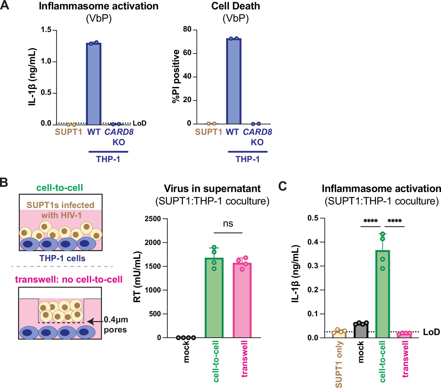
Our previous work investigating HIV-dependent CARD8 inflammasome activation used the cationic polymer DEAE-dextran, which is a common reagent used to enhance viral infection in cell culture (Bailey et al., 1984). However, we found that DEAE-dextran could induce inflammasome activation in the absence of viral infection in some ‘wildtype’ (WT) THP-1 cell stocks (see Appendix 1). These results prompted us to establish other models of HIV-1 infection and subsequent inflammasome activation that lack cationic polymers. Thus, we designed an in vitro coculture infection system to mimic HIV-1 cell-to-cell transmission by infecting SUPT1 cells, a T-cell lymphoma line (i.e., donor cells) and then mixing them with uninfected THP-1 cells (i.e., target cells). We opted for SUPT1 cells as the viral producer cell line because they are permissive to HIV-1 infection, and unlike THP-1 cells, SUPT1 cells do not respond to a known CARD8 inflammasome activator, ValboroPro (VbP), as assayed by both IL-1β secretion and cell death, indicating that SUPT1 cells do not have a functional CARD8 inflammasome pathway (Figure 1A). This allowed us to infer that inflammasome outputs (e.g., IL-1β secretion) in our coculture system occur upon cell-to-cell transmission of HIV-1 from SUPT1 cells to the CARD8-competent THP-1 cells.
Figure 1
HIV-1 cell-to-cell infection induces inflammasome activation.
(A) SUPT1 or THP-1 cells were primed with Pam3CSK4 (500ng/mL) overnight then treated with 5 μM ValboroPro (VbP) for 24 hours, then assessed for IL-1β secretion and cell death via propidium iodide (PI) uptake. %PI positive was normalized to mock-infected controls. (B) (left) Schematic illustrating the experimental setup for SUPT1:THP-1 cell coculture either with (bottom) or without (top) a transwell. (right) SUPT1 cells were either mock-infected or infected with HIV-1LAI then cocultured with primed WT THP-1 cells (see Methods) 20 hours post infection. Mock- or HIV-1LAI-infected SUPT1 cells were either mixed with the THP-1 cells or put in a transwell with a virus-permeable membrane as shown in panel (B) (left). Supernatant in the cell-to-cell condition and in the supernatant outside of the transwell were sampled and measured for released HIV virions via a reverse transcriptase (RT) assay or (C) IL-1β secretion 3 days after starting the coculture. Dotted line indicates limit of detection (LoD). Datasets represent mean ± SD (A: n=2; B, C n=4 biological replicates). One-way ANOVA with (B) Tukey’s or (C) Dunnett’s test using GraphPad Prism 10. ns = not significant, *p<0.05, **p<0.01, ***p<0.001, ****p<0.0001.
We found that coculture of THP-1 cells with HIV-1LAI-infected SUPT1 cells (Figure 1B) but not mock-infected SUPT1 cells results in robust inflammasome activation as indicated by IL-1β secretion, suggesting that our coculture system, which lacks DEAE-dextran, can induce HIV-dependent inflammasome activation via cell-to-cell infection (Figure 1C). To further test this assumption, we prevented cell-to-cell contact using a virus-permeable transwell with a 0.4 μm pore insert (Figure 1B, left). We verified that there were equivalent amounts of infectious virus in the cell-to-cell condition versus the lower chamber of the transwell condition by measuring RT activity in the supernatant (Figure 1B, right). Despite equivalent amounts of infectious virus in both conditions, we observed that HIV-1LAI-infected SUPT1 cells (upper chamber) cocultured with THP-1 cells (lower chamber) did not lead to detectable IL-1β secretion (Figure 1C). These data suggest that our SUPT1:THP-1 coculture system can trigger HIV-dependent inflammasome activation in a manner dependent on cell-to-cell contact.
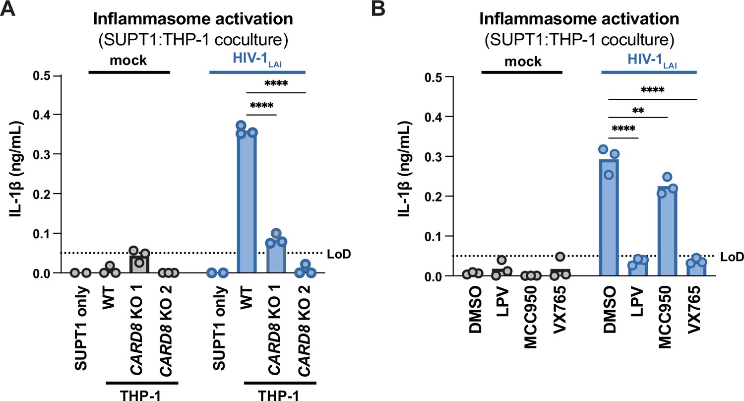
We next assessed the role of CARD8 and other inflammasome sensors during cell-to-cell transmission of HIV-1. We cocultured mock- or HIV-1LAI-infected SUPT1 cells with either WT or CARD8 KO THP-1 cells and compared inflammasome activation by measuring levels of secreted IL-1β. HIV-1LAI-infected SUPT1 cells cocultured with WT but not CARD8 KO THP-1 cells resulted in a significant increase in IL-1β (Figure 2A). These results suggest that CARD8 is the primary sensor that drives inflammasome activation in HIV-1 cell-to-cell transmission to THP-1 cells. Since the NLRP3 inflammasome has previously been implicated in HIV-dependent inflammasome activation (Bandera et al., 2018; Chivero et al., 2017; Hernandez et al., 2014; Leal et al., 2020; Mamik et al., 2017; Zhang et al., 2021), we also assessed the effects of the NLRP3 inflammasome-specific inhibitor MCC950 (Coll et al., 2015; Primiano et al., 2016) on inflammasome activation in our coculture system. Treatment with MCC950 or the caspase 1 (CASP1) inhibitor VX765 (Wannamaker et al., 2007) were sufficient to abrogate inflammasome activation induced by the ionophore nigericin, a well-characterized NLRP3 agonist (Figure 2—figure supplement 1A). However, in the HIV-1 coculture system, MCC950 treatment had only a modest effect on inflammasome activation while VX765 and the HIVPR inhibitor lopinavir (LPV), which prevents CARD8 cleavage by HIV-1PR (Kulsuptrakul et al., 2023; Wang et al., 2021), completely abrogated IL-1β secretion (Figure 2B). We observed similar results during HIV-1LAI and HIV-1LAI-VSVG cell-free infection of THP-1 cells in the presence of DEAE-dextran (Figure 2—figure supplement 1B). Taken together, these findings indicate that HIV-dependent inflammasome activation via cell-to-cell transmission is CARD8-dependent and largely NLRP3-independent.
Figure 2 with 1 supplement see all
HIV-1 cell-to-cell transmission induces CARD8-dependent activation largely independent of NLRP3.
(A) SUPT1 cells were either mock-infected or infected with HIV-1LAI for 18–20 hours prior to coculture with wildtype (WT) or CARD8 knockout (KO) THP-1 cells. The coculture was harvested 72 hours later to probe for IL-1β secretion in the coculture supernatant via IL-1R reporter assay. THP-1 cells were primed with Pam3CSK4 (500ng/mL) for 16–24 hours prior to coculture. SUPT1 cells were infected with HIV-1LAI such that 30% of the cells were positive for intracellular p24gag after 18–20 hours. (B) SUPT1 cells were either mock- or HIV-1LAI-infected as in (A) for 18–20 hours then incubated in DMSO, lopinavir (LPV), MCC950, or VX765 at 0.01%, 5 μM, 10μM, or 1μg/mL, respectively, for 15 minutes prior to coculturing with primed WT THP-1 cells. The coculture was assessed for subsequent inflammasome activation after 72 hours as in (A). Dotted line indicates limit of detection (LoD). Datasets represent mean ± SD (n=3 biological replicates). Two-way ANOVA with Dunnett’s test using GraphPad Prism 10. ns = not significant, *p<0.05, **p<0.01, ***p<0.001, ****p<0.0001.
CARD8, but not NLRP3, is required for inflammasome activation during HIV-1 cell-to-cell transmission into primary monocyte-derived macrophages
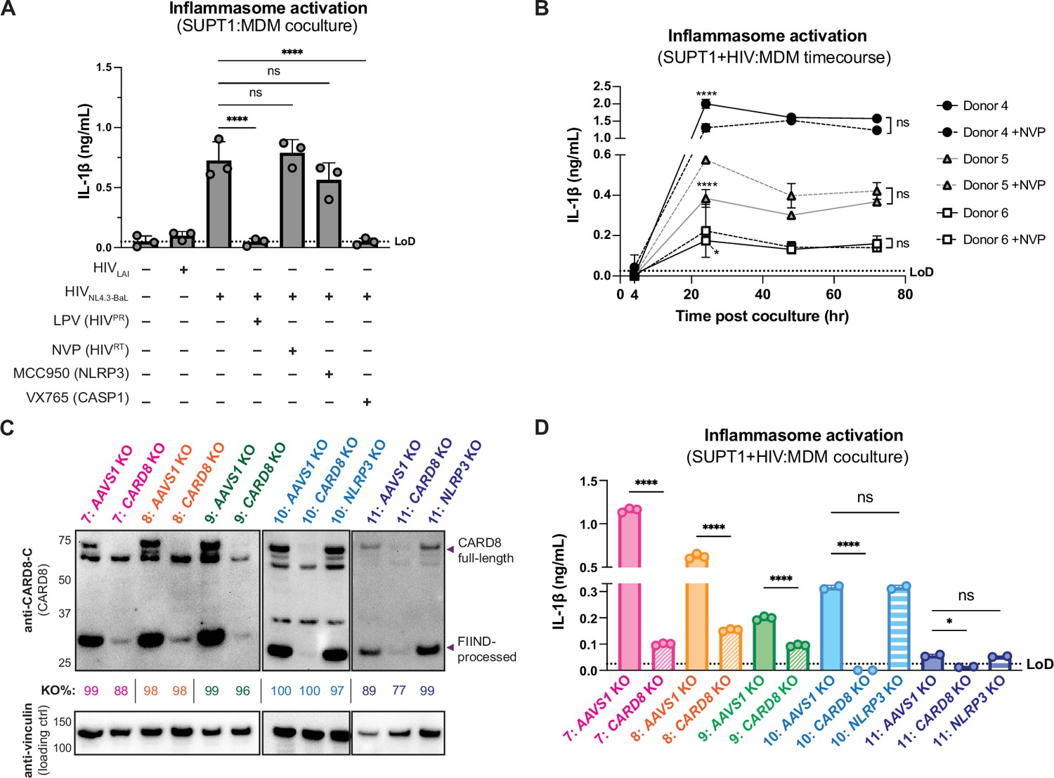
We next examined inflammasome activation upon HIV-1 cell-to-cell transmission in primary human monocyte-derived macrophages (MDMs). Previously, we had observed that CARD8 could sense active HIV-1PR released into the host cytosol following viral fusion, which we refer to as ‘incoming’ HIV-1PR in our cell-free infection system in THP-1 cells using DEAE-dextran and spinoculation (Kulsuptrakul et al., 2023). Thus, we assessed the importance of viral entry by coculturing MDMs from three independent donors with mock-, HIV-1LAI-, or HIV-1NL4.3-BaL-infected SUPT1 cells expressing CCR5 (SUPT1-CCR5) and assayed for inflammasome activation (Figure 3A). HIV-1LAI is a CXCR4 tropic strain unable to infect macrophages, whereas HIV-1NL4.3-BaL uses CCR5 as a co-receptor which is required for infection of macrophages. We observed inflammasome activation, as measured by IL-1β secretion, in MDMs cocultured with HIV-1NL4.3-BaL-infected SUPT1 cells but not in MDMs cocultured with mock- or HIV-1LAI-infected SUPT1-CCR5 cells (Figure 3A). This demonstrates that HIV-dependent inflammasome activation can occur in MDMs during cell-to-cell infection in a manner dependent on viral entry. To further ascertain if this inflammasome activation was CARD8-dependent and driven by incoming HIVPR during SUPT1:MDM cell-to-cell transmission, we investigated the effects of different inhibitors on inflammasome activation in MDM cocultures with HIV-1NL4.3-BaL-infected SUPT1s. We observed that IL-1β secretion was abrogated by treatment with lopinavir, an HIV-1 protease inhibitor, and VX765, a CASP1 inhibitor, indicating that inflammasome activation in MDM cocultures is dependent on HIVPR and CASP1, respectively (Figure 3A). In addition, we used an RT inhibitor, nevirapine (NVP), to prevent synthesis of de novo translated HIVPR, and thus any CARD8-dependent IL-1β secretion would only be due to incoming HIVPR in the presence of NVP. Indeed, we observed HIV-dependent inflammasome activation in the presence of NVP that was added at the time of coculture, indicating that incoming HIVPR is sufficient to elicit an inflammasome response (Figure 3A). Lastly, MDM cocultures treated with MCC950, an inhibitor of the NLRP3 inflammasome, had no effect on IL-1β secretion (Figure 3A), and we observed similar inflammasome activation results regardless of whether the MDMs were primed with TLR1/2 agonist Pam3CSK4 versus TLR4 agonist lipopolysaccharide (LPS) (Figure 3—figure supplement 1A). Thus, cell-to-cell contact of infected cells with primary monocyte-derived macrophages can elicit an inflammasome response in a manner that is dependent on viral entry, CASP1, and incoming HIVPR, and independent from NLRP3.
Figure 3 with 1 supplement see all
Cell-to-cell HIV infection induces CARD8-dependent inflammasome activation in monocyte-derived macrophages (MDMs).
(A) MDMs from three independent donors were cocultured with SUPT1 cells expressing CCR5 (SUPT1-CCR5) that were mock-, HIV-1LAI-, or HIV-1NL4.3-BaL-infected then assayed for inflammasome activation 48 hours post coculture for IL-1β secretion. Fifteen minutes before starting the coculture, SUPT1-CCR5 cells infected with HIV-1NL4.3-BaL were pre-treated with either DMSO, lopinavir (5μM), nevirapine (50 μM), MCC950 (10 μM), or VX765 (1μg/mL), inhibiting HIV-1 protease (HIVPR), HIV-1 reverse transcriptase (HIVRT), NLRP3, or caspase 1 (CASP1), respectively. (B) MDMs from three independent donors were cocultured with SUPT1-CCR5 cells infected with HIV-1NL4.3-BaL in either the presence or absence of nevirapine (NVP). Supernatant was harvested at 4, 24, 48, or 72 hours to assay for IL-1β secretion. (C) MDMs from five independent donors were knocked out (KO) for AAVS1 CARD8, or NLRP3 using a Synthego gene KO kit then immunoblotted using an anti-CARD8 antibody or anti-vinculin. Full-length and FIIND-processed CARD8 intermediates are marked with a purple arrow. Size is indicated in kDa on left side of blot. Table between CARD8 and vinculin blot shows Synthego gene KO% scores for each donor KO line. (D) AAVS1, CARD8 or NLRP3 KO MDM lines from (C) were primed with Pam3CSK4 (500 ng/mL) overnight and then cocultured with SUPT1-CCR5 cells mock-, or HIV-1NL4.3-BaL-infected then assayed for inflammasome activation 48 hours post coculture for IL-1β secretion. For all SUPT1:MDM experiments, SUPT1-CCR5 cells were infected with HIV-1LAI or HIV-1NL4.3-BaL such that 5–20% of cells were positive for intracellular p24gag after 20 hours. IL-1 levels shown were normalized to the SUPT1 mock-infected coculture control. Dotted line indicates limit of detection (LoD). Datasets represent mean ± SD (A: n=3 independent donors, B: n=2 biological replicates for each donor, D: n=3 technical replicates per donor). One-way ANOVA with (A) Tukey’s or (D) Sidak’s test or (B) two-way ANOVA with Tukey’s test using GraphPad Prism 10. ns = not significant, *p<0.05, **p<0.01, ***p<0.001, ****p<0.0001.
-
Figure 3—source data 1
Original western blot images used to generate Figure 3C.
- https://cdn.elifesciences.org/articles/102676/elife-102676-fig3-data1-v1.pdf
-
Figure 3—source data 2
Original western blot .tif files used to generate Figure 3C.
- https://cdn.elifesciences.org/articles/102676/elife-102676-fig3-data2-v1.zip
To further assess the timing of this inflammasome activation, we conducted a SUPT1:MDM time-course coculture experiment with three additional donors, assaying IL-1β secretion at 4, 24, 48, and 72 hours post coculture in the presence or absence of NVP. We observed inflammasome activation as determined by IL-1β secretion by 24 hours post coculture in all donors that persisted at a similar level at 48 and 72 hours post coculture (Figure 3B). As expected, we observed donor-to-donor variation in the extent to which IL-1β secretion occurred following HIV-1 infection. However, per donor, HIV-1-driven IL-1β levels were comparable to VbP-induced inflammasome activation (Figure 3—figure supplement 1B). Moreover, adding NVP had no effect on IL-1β secretion in HIV-1-infected MDM cocultures (Figure 3B). To verify that NVP was functional, we assayed the supernatant of the mock- and NVP-treated cocultures from Figure 3B at 48 hours post coculture for infectious virions via an assay that measures RT activity and observed lower RT activity in NVP-treated MDM donors (Figure 3—figure supplement 1C). Unlike the ‘cell-free’ infection conditions in which we previously observed an increase in IL-1β levels 4 hours post-infection, we did not detect measurable differences in IL-1β secretion at this early timepoint following the establishment of SUPT1:MDM coculture. Nevertheless, the data are consistent with incoming HIVPR being responsible for inflammasome activation during cell-to-cell transmission of HIV because the induction of IL-1β persists in the presence of NVP which would block any de novo synthesis of new gag/pol products (Figure 3A and B). Taken together, these data suggest that in the context of cell-to-cell transmission, CARD8 is likely the inflammasome-forming sensor that detects HIV-1 infection via incoming HIVPR activity in primary monocyte-derived macrophages.
To specifically address the role of CARD8 in HIV-1-induced inflammasome activation in MDMs, we genetically edited MDMs by isolating monocytes from five donors and electroporated them with Cas9 RNPs complexed with three unique sgRNAs per gene targeting AAVS1, a safe harbor locus, CARD8 or NLRP3 (only two donors for NLRP3 KO). Edited MDMs were then differentiated for 6 days prior to evaluating KO efficiency and initiating cocultures with HIV-1-infected SUPT1 cells. To verify KO efficiency, we immunoblotted with an antibody that detects the CARD8 C-terminus in CARD8 KO MDMs relative to the AAVS1 KO control and observed a marked reduction of the full-length and FIIND-processed CARD8 protein in all five donors (Figure 3C). In addition, we confirmed AAVS1, CARD8, and NLRP3 KO at the genetic level via Synthego ICE analysis (Conant et al., 2022), measuring >85% KO efficiency (Figure 3C). We also observed robust inflammasome activation upon treatment with CARD8 inflammasome activator VbP as measured by IL-1β secretion in AAVS1 KO MDMs from two of the three donors, which was completely abrogated in CARD8 KO MDMs, confirming functional loss of CARD8 (Figure 3—figure supplement 1D). We then cocultured either AAVS1 KO, CARD8 KO, or NLRP3 KO MDMs with mock- or HIV-1NL4.3-BaL-infected SUPT1-CCR5 cells at a 1:1 ratio and measured inflammasome activation via IL-1β secretion 48 hours post coculture. In all five donors, we observed significant reduction in inflammasome activation in CARD8 KO cocultures relative to the AAVS1 KO control (Figure 3D). Consistent with our findings with SUPT1:THP-1 coculture, we observed no difference in inflammasome activation when coculturing infected SUPT1 cells with AAVS1 KO vs NLRP3 KO MDMs, suggesting that the inflammasome activation was also largely NLRP3-independent (Figure 3D). Taken together, these data demonstrate that CARD8 is required for inflammasome activation in MDMs during HIV-1 cell-to cell transmission.
Coculture of HIV-1-infected primary CD4+ T cells with primary MDMs elicits CARD8-dependent inflammasome activation
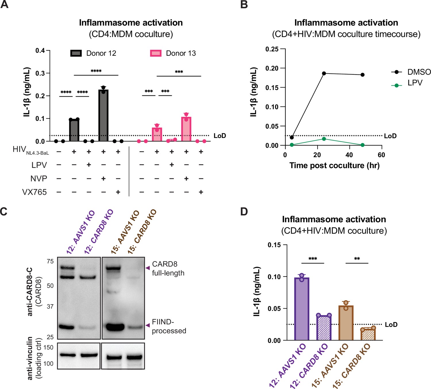
We next investigated inflammasome activation in the context of cell-to-cell infection using primary CD4+ T cells as donor cells, rather than SUPT1 cells, and primary MDMs as target cells. Mock- or HIV-1NL4.3-BaL-infected CD4+ T cells were cocultured with MDMs in the presence or absence of various inhibitors (as in Figure 3A). We assessed inflammasome activation via IL-1β secretion 72 hours post coculture and observed inflammasome activation when coculturing with HIV-infected T cells but not mock-infected T cells or cocultures treated with LPV or VX765, demonstrating that inflammasome activation is driven by HIVPR and CASP1 (Figure 4A) and consistent with our findings using the SUPT1:MDM coculture (Figure 3). We also observed that inflammasome activation persisted in NVP-treated cocultures, suggesting that incoming protease is also important for inflammasome activation in the context of cell-to-cell transmission of HIV-1 from primary CD4+ T cells to primary MDMs (Figure 4A). To confirm the potency of LPV and NVP, we assayed for infectious virions in the supernatant of these CD4:MDM cocultures and detected a dramatic decrease in RT activity in the presence of either of these drugs, indicating that these drugs were efficacious at this dose in these cells (Figure 4—figure supplement 1A). We also conducted a time-course experiment with a CD4:MDM coculture from an independent donor in the presence or absence of LPV. Similar to the SUPT1:MDM time course (Figure 3B), we were able to detect elevated levels of IL-1β by 24 hours post coculture, which was again strictly dependent on the enzymatic activity of the viral protease as LPV treatment completely inhibited IL-1β secretion (Figure 4B).
Figure 4 with 1 supplement see all
Primary CD4+ T cell:MDM coculture elicits CARD8-dependent inflammasome activation.
(A) CD4+ T cells from a blood donor were isolated, activated, and either mock-infected or infected with HIV-1NL4.3-BaL for 3 days such that ~10% of cells were positive for intracellular p24gag. Monocyte-derived macrophages (MDMs) were primed with Pam3CSK4 then cocultured with mock- or HIV-1-infected primary CD4 T cells in the presence or absence of lopinavir (LPV), nevirapine (NVP), or VX765, inhibiting HIV protease, reverse transcriptase, or caspase 1, respectively. Supernatants were harvested 3 days post coculture to assay for IL-1β secretion via IL-1 reporter assay. (B) CD4+ T cells from donor 12 and MDMs from donor 14 were cocultured as in (A) in the presence or absence of LPV. Supernatant was harvested at 4, 24 and 48 hours post coculture to probe for IL-1β secretion. (C) AAVS1 or CARD8 MDM KOs were immunoblotted using an anti-CARD8 antibody or anti-vinculin. Full-length and FIIND-processed CARD8 intermediates are marked with a purple arrow. Size is indicated in kDa on left side of blot. (D) AAVS1 or CARD8 KO MDMs from (C) were cocultured with CD4+ T cells infected with HIV-1NL4.3-BaL then assayed for IL-1β secretion 48 hours post coculture. The donor 12 cocultures consisted of autologous CD4s and MDMs, whereas the MDMs from donors 13–15 were cocultured with donor 12 CD4s. Dotted line indicates limit of detection (LoD). Datasets represent mean ± SD (A, D: n=2 technical replicates for each donor, B: n=3 technical replicates for one donor). (A) Two-way ANOVA with Tukey’s test (D) One-way ANOVA with Sidak’s test using GraphPad Prism 10. ns = not significant, *p<0.05, **p<0.01, ***p<0.001, ****p<0.0001.
-
Figure 4—source data 1
Original western blot images used to generate Figure 4C.
- https://cdn.elifesciences.org/articles/102676/elife-102676-fig4-data1-v1.pdf
-
Figure 4—source data 2
Original western blot .tif files used to generate Figure 4C.
- https://cdn.elifesciences.org/articles/102676/elife-102676-fig4-data2-v1.zip
To interrogate the specific role of CARD8 in this primary CD4:MDM coculture system, we generated AAVS1 KO (as a control) or CARD8 KO MDMs and cocultured the MDMs with either mock- or HIV-infected primary CD4+ T cells then assayed for inflammasome activation 48 hours post coculture. KO efficiency was confirmed via immunoblot and functional response to VbP (in one of the two donors) (Figure 4C, Figure 4—figure supplement 1B). We detected a significant decrease in inflammasome activation when infected T cells were cocultured with CARD8 KO versus AAVS1 KO MDMs (Figure 4D), indicating that CARD8 is required for inflammasome activation in MDMs during HIV-1 transmission from primary CD4+ T cells to MDMs. Taken together, our data indicate that CARD8 plays a pivotal role in sensing and responding to HIV-1 cell-to-cell infection between primary CD4 T cells and macrophages.
Protease inhibitor-resistant strains of HIV-1 differentially cleave and activate CARD8
The consequences of CARD8 inflammasome activation on viral replication have been challenging to assess given that viral fitness is intrinsically linked to viral protease processing of the viral polyprotein such that inhibiting HIVPR also prevents viral replication. In an attempt to circumvent this issue, we surveyed a panel of multi-HIVPR inhibitor-resistant (PI-R) infectious molecular clones of HIV-1 (Varghese et al., 2013). This panel of PI-R molecular clones varies in resistance to HIV protease inhibitors, including nelfinavir (NFV), fosamprenavir (FPV), saquinavir (SQV), indinavir (IDV), atazanavir (ATV), lopinavir (LPV), tipranavir (TPV), and darunavir (DRV). Each molecular clone encodes 4–11 mutations in HIVPR as well as various compensatory HIVgag mutations (Varghese et al., 2013; Supplementary file 1).
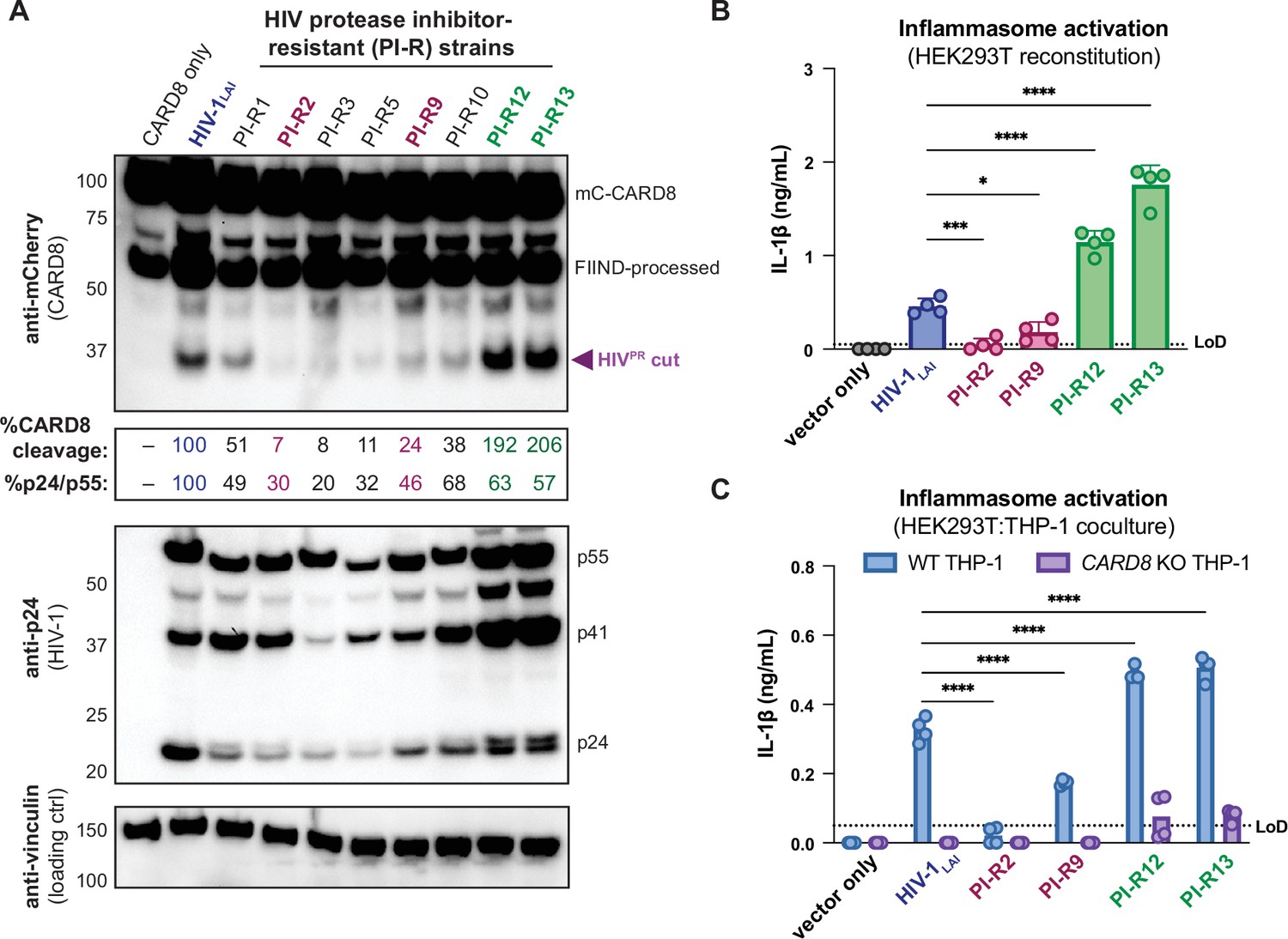
We initially tested if PI-R HIV-1 proviruses differentially cleave CARD8 by co-transfecting HEK293T cells with an expression plasmid encoding an N-terminal mCherry tagged human CARD8 and either wildtype HIV-1LAI or PI-R HIV-1 proviruses. HIV-1LAI protease cleaves CARD8 between phenylalanine (F) 59 and F60 (Wang et al., 2021), resulting in an ~33 kDa product (Figure 5A, top). By quantifying the 33 kDa CARD8 cleavage product with each HIV-1 provirus, we identified a PI-R clone that exhibited similar efficiency at cleaving CARD8 to HIV-1LAI (i.e., PI-R1), PI-R clones that were markedly less efficient at cleaving CARD8 than HIV-1LAI (i.e., PI-R2, PI-R3, PI-R5, PI-R9, and PI-R10) and two PI-R clones, PI-R12 and PI-R13, that were more efficient at cleaving CARD8 than HIV-1LAI (Figure 5A, top, Supplementary file 1). Of note, all PI-R proviruses had similar levels of HIVPR activity for HIVgagpol polyprotein processing from p55gag to p24gag as indicated by the ratio of p24gag/p55gag quantified from the anti-p24gag immunoblot (Figure 5A, middle). These results indicate that naturally occurring HIV-1 protease mutations can influence host targets like CARD8.
Figure 5
Protease inhibitor-resistant strains of HIV-1 differentially cleave and activate CARD8.
(A) HEK293T cells were transfected with a construct encoding CARD8 with an N-terminal mCherry tag (mCherry-CARD8) and indicated HIV-1 proviral constructs. Protease inhibitor-resistant (PI-R) clones of HIV-1 are a subset of a panel expressing prototypical multidrug resistant HIV-1 protease (HIVPR) in an NL4.3 backbone (Supplementary file 1). Top: immunoblotting using anti-mCherry antibody to detect mCherry-CARD8. The full-length (mC-CARD8) and FIIND-processed bands are indicated as well as the HIVPR cut product. Size is indicated in kDa on left side of blot. The band at ~45 kDa is the result of cleavage by the 20S proteasome (Hsiao et al., 2022). % CARD8 cleavage was calculated by quantifying the HIVPR cut band relative to the HIV-1LAI control using BioRad Image Lab 6. Middle: immunoblotting with an anti-p24gag antibody showing HIV-1gag cleavage products p41gag and p24gag, and/or full-length HIV-1gag, p55gag. %p24/p55 was calculated from the ratio of p24gag versus p55gag product by quantifying the volume of the p24gag bands versus the p55gag band relative to the HIV-1LAI control using BioRad Image lab 6. Bottom: immunoblotting with an anti-vinculin antibody to detect vinculin as a loading control. (B) HEK293T cells were transfected with human caspase 1 and human pro-IL-1β, and either carrier vector or indicated HIV-1 proviruses then probed for IL-1β secretion 24 hours post-transfection via IL-1R reporter assay. (C) HEK293T cells were transfected with indicated HIV-1 proviruses (300ng). 24 hours post-transfection either wildtype (WT) or CARD8 knockout (KO) THP-1s were overlayed on the transfected HEK293T cells in a 1:1 ratio. THP-1s were primed with Pam3CSK4 overnight prior to coculture. Supernatants were harvested 24 hours post coculture to assay for IL-1β secretion as in (B). Dotted line indicates limit of detection (LoD). Datasets represent mean ± SD (n=4 biological replicates). p-Values were determined by two-way ANOVA with Dunnett’s test using GraphPad Prism 10. ns = not significant, *p<0.05, **p<0.01, ***p<0.001, ****p<0.0001.
-
Figure 5—source data 1
Original western blot images used to generate Figure 5A.
- https://cdn.elifesciences.org/articles/102676/elife-102676-fig5-data1-v1.pdf
-
Figure 5—source data 2
Original western blot .tif files used to generate Figure 5A.
- https://cdn.elifesciences.org/articles/102676/elife-102676-fig5-data2-v1.zip
We next assessed if PI-R clones exhibiting reduced (PI-R2 and -9) or increased (PI-R12 and -13) cleavage of CARD8 relative to HIV-1LAI (Figure 5A and B, Supplementary file 1) resulted in differential inflammasome activation. HEK293T cells endogenously express CARD8 but lack the downstream components (i.e., CASP1, GSDMD, and IL-1β/IL18) of the inflammasome pathway. Thus, we reconstituted the inflammasome pathway in HEK293T cells by co-transfection of human caspase 1, human pro-IL-1β, and either empty vector, HIV-1LAI or representative PI-R proviruses then quantified CASP1-dependent processing of pro-IL-1β as a readout of CARD8 inflammasome activation as in Tsu et al., 2023. Consistent with the observed differences in CARD8 cleavage by PI-R clones (Figure 5A), we found that PI-R2 and PI-R9, which exhibited less CARD8 cleavage than HIV-1LAI, also induced lower IL-1β levels than HIV-1LAI (Figure 5B). Similarly, PI-R12 and PI-R13, which demonstrated enhanced CARD8 cleavage, elicited higher IL-1β levels than HIV-1LAI (Figure 5B). However, these PI-R clones, relative to the LAI strain, may have distinct protease substrate specificity, variable efficiency/kinetics in viral assembly, gag dimerization, and other factors, which may also influence CARD8 inflammasome activation. We next assessed inflammasome activation by the PI-R clones in a cell-to-cell transmission model using HEK293T cells as donor cells rather than SUPT1 cells and either WT or CARD8 KO THP-1 cells as the target line at a 1:1 ratio. We opted to overexpress the HIV-1LAI or the PI-R proviruses in HEK293T cells rather than infecting SUPT1 cells due to dramatic variability in replication kinetics between PI-R strains. In these HEK293T:THP-1 cocultures, we observed that cell-to-cell transmission of PI-R2 and PI-R9 resulted in lower IL-1β levels while PI-R12 and PI-R13 resulted in higher IL-1β levels compared to HIV-1LAI, respectively (Figure 5C), consistent with our findings from CARD8 cleavage (Figure 5A) and reconstituted inflammasome assays (Figure 5B). Our findings suggest that HIV-dependent inflammasome activation is under genetic control of the viral protease in a manner that can be increased or decreased with naturally occurring mutations induced by drug resistance.
Discussion
We demonstrate that cell-to-cell transmission of HIV-1 from T cells to myeloid cells in both immortalized and primary cell coculture models of infection yields CARD8-dependent inflammasome activation via incoming HIVPR. This inflammasome activation occurs in a largely NLRP3-independent manner. In addition, we identified protease inhibitor-resistant strains of HIV-1 that differentially cleave and activate the CARD8 inflammasome. Thus, HIVPR mutants selected for their resistance to different protease inhibitors also affect their ability to cleave host proteins including the inflammasome-forming sensor CARD8.
CARD8 is the primary inflammasome-forming sensor of HIV-1 infection
Previously, both the NLRP3 and IFI16 inflammasomes had been implicated as innate sensors of HIV-1 infection and drivers of CD4+ T cell depletion using blood and lymphoid-derived CD4+ T cells, respectively, and cell-to-cell transmission was reported to be crucial for IFI16 sensing of abortive HIV transcripts (Doitsh et al., 2014; Galloway et al., 2015; Monroe et al., 2014; Zhang et al., 2021). However, the mechanism of NLRP3 inflammasome activation in response to HIV-1 remains elusive. Similarly, there have been reports that IFI16 is not an inflammasome-forming sensor, and instead a nuclear transcriptional regulator of antiviral genes including type I interferons and RIG-I (Hornung et al., 2009; Jiang et al., 2021; Thompson et al., 2014), suggesting that there may be other mechanisms of CD4+ T cell depletion and HIV-dependent inflammasome activation at play. Indeed, CARD8, which is expressed and functional in naïve and memory CD4+ and CD8+ T cells (Linder et al., 2020), was recently shown to be required for pyroptosis in primary human blood- and lymphoid-derived CD4+ T cells and humanized mouse models (Wang et al., 2024), implicating CARD8 as a major driver of CD4+ T cell depletion during HIV-1 infection. In this study and our prior work (Kulsuptrakul et al., 2023), we demonstrate that CARD8 is also the primary innate sensor during HIV-1 infection in myeloid cell types during cell-to-cell transmission. However, our present study does not rule out the possibility that under certain conditions or in certain cell types, NLRP3 inflammasome activation may occur, for example, following GSDMD pore formation following CARD8 inflammasome activation and play a more profound role in promoting HIV-dependent inflammation. Nonetheless, these data, along with other recent work (Wang et al., 2024), strongly suggest that CARD8 is a major innate sensor of HIV-1 infection.
Protease inhibitor resistance mutations and inflammatory disease
Given the important role of HIVPR in replication, early combination antiretroviral therapy for people living with HIV (PLWH) included protease inhibitors along with RT inhibitors. However, resistance mutations to protease inhibitors arose in PLWH through mutations around the HIVPR active site allowing for polyprotein processing and viral maturation while avoiding drug inhibition. Despite typically having poor overall viral fitness due to less efficient polyprotein processing and replication relative to wildtype HIV-1 in the absence of protease inhibitors, these mutant drug-resistant HIV-1 strains can persist in PLWH on antiviral therapy (Luca, 2006; Martinez-Picado et al., 1999; Prado et al., 2002; Resch et al., 2002). To compensate for mutations in HIVPR that change its substrate specificity, HIVgag sometimes evolves mutations around HIVPR cleavage sites to permit proper polyprotein processing (Varghese et al., 2013). Here, we identified multiple HIVPR inhibitor-resistant strains of HIV-1 that can differentially cleave and activate the CARD8 inflammasome (Figure 5, Supplementary file 1). As the degree of inflammation is a better predictor of disease progression in untreated individuals than viral load (Deeks et al., 2004; Giorgi et al., 1999), we speculate that differential CARD8 inflammasome activation could influence disease progression for PLWH harboring HIVPR resistance mutations that cleave CARD8 more or less efficiently. More broadly, we suggest that host targets of viral proteases like CARD8 may influence the selection of viral variants during treatment with antiviral protease inhibitor monotherapies.
Viral protease influx activates the CARD8 inflammasome
In this study, we found that HIV-dependent CARD8 inflammasome activation during cell-free infection requires a cationic polymer like DEAE-dextran to facilitate efficient viral infection (see Appendix 1). Despite being infected with the same amount of virus and exhibiting similar percent infection 24 hours post-infection, as measured by intracellular p24gag, with and without DEAE-dextran, we hypothesize that DEAE-dextran during cell-free infection may increase the total viral dose that enters cells. This increased viral dose in the presence of DEAE-dextran could consist of both infectious particles and non-infectious particles that may nonetheless contain active protease, leading to more efficient viral protease influx to trigger CARD8 sensing. Hence, the percentage of p24gag-positive cells after 24 hours may be an underestimate of the total amount of viral entry in the DEAE-dextran condition. We speculate that a considerable influx of incoming HIVPR may be necessary to induce CARD8 inflammasome activation.
We also observed that DEAE-dextran can trigger inflammasome activation in some THP-1 cell lines (see Appendix 1), prompting us to assay for inflammasome activation upon HIV-1 cell-to-cell transmission with target cells at a 1:1 ratio in the absence of cationic polymer. We found that inflammasome activation following cell-to-cell transmission of HIV-1 could be detected by 24 hours (Figure 3B), which is delayed relative to our detection of CARD8 inflammasome activation 2 hours post cell-free HIV-1 infection in the presence of DEAE-dextran (Kulsuptrakul et al., 2023). Nonetheless, we still hypothesize that this inflammasome activation is driven by active incoming viral protease because treatment with an RT inhibitor has no effect on inflammasome activation, which implies that de novo protease production is not necessary (Figures 3A, B, 4A, B). Similarly, we observed that IL-1β levels do not increase after plateauing 24 hours after establishing the coculture (Figures 3B and 4B), suggesting that secondary infection does not further amplify inflammasome activation. We infer that this is also likely a product of the efficiency of viral entry and the necessity for multiple virions infecting at the same time to deliver a sufficient amount of active HIVPR for cytosolic CARD8 sensing. We postulate that under certain physiological conditions, cell-to-cell transmission can cause CARD8 inflammasome activation when there is an influx of active incoming HIVPR across the viral synapse. Taken together, we speculate that both cell-free infection facilitated by cationic polymer and cell-to-cell transmission can achieve sufficient levels of active HIVPR influx to activate the CARD8 inflammasome.
Macrophages have been reported to be primarily infected through phagocytosis of infected CD4+ T cells or cell-to-cell transmission (Dupont and Sattentau, 2020; Martínez-Méndez et al., 2017; Orenstein, 2000). We demonstrate that unlike CD4+ T cells, which are rapidly depleted by HIV-1 infection and do not release IL-1β or IL-18 (Linder et al., 2020), primary macrophages release pro-inflammatory cytokines in response to HIVPR during cell-to-cell infection from infected primary CD4+ T cells and T cell lines (Figures 3 and 4), thus representing a potential source of sustained IL-1β and subsequent chronic immune activation. In addition to promoting chronic immune activation, HIV-dependent IL-1β release from macrophages may also contribute to HIV-1 pathogenesis by activating nearby CD4+ T cells, rendering them susceptible to becoming infected with HIV-1, and thus indirectly promoting CD4+ T cell depletion. Collectively with our prior work (Kulsuptrakul et al., 2023), our findings provide further evidence that CARD8 inflammasome activation is driven by incoming HIVPR under conditions where multiple virions may enter cells, and thus could be a potential driver of HIV-1 pathogenesis by promoting chronic immune activation.
Materials and methods
Plasmids and reagents
Request a detailed protocolpMD2.G used for HIV-1LAI-VSVG production was a gift from Didier Trono (Addgene). HIV-1LAI has been previously described (Peden et al., 1991). The following reagents were obtained through the NIH HIV Reagent Program, Division of AIDS, NIAID, NIH: lopinavir (LPV), nevirapine (NVP), human immunodeficiency virus 1 (HIV-1) NL4-3 BaL Infectious Molecular Clone (p20-36) (HIV-1NL4.3-BaL), ARP-11442, contributed by Dr. Bruce Chesebro (Chesebro et al., 1992; Chesebro et al., 1991; Toohey et al., 1995; Walter et al., 2005), and Panel of Multi-Protease Inhibitor Resistant Infectious Molecular Clones, HRP-12740, contributed by Dr. Robert Shafer (Varghese et al., 2013). Mutant HIVPR sequences were amplified from clinically derived viral cDNA encoding protease genes with resistance to multiple protease inhibitors then cloned into an NL4.3 backbone with overhangs including the 3’ end of gag with the gag cleavage site and the 5’ end of RT as previously described (Varghese et al., 2013). CARD8 variant constructs were cloned as previously described (Kulsuptrakul et al., 2023). VX765 and MCC950 were sourced from Invivogen (cat: inh-vx765i-1 and inh-mcc, respectively).
Cell culture
Request a detailed protocolSUPT1 (ATCC) and THP-1 cells (JK and ATCC) were cultured in RPMI (Invitrogen) with 10% FBS, 1% penicillin/streptomycin antibiotics, 10 mM HEPES, 0.11 g/L sodium pyruvate, 4.5 g/L d-glucose, and 1% GlutaMAX. JK THP-1 cells were used for all experiments in this manuscript and our previous work unless explicitly stated (see Appendix 1; Kulsuptrakul et al., 2023). Primary monocytes were cultured in RPMI (Invitrogen) with 10% FBS, and 1% penicillin/streptomycin antibiotics and differentiated in the presence of 20 ng/mL GM-CSF (Peprotech cat: 300-03) and 20 ng/mL M-CSF (Peprotech cat: 300-25). Primary CD4+ T cells were cultured in RPMI (Invitrogen) with 10% FBS, 1% penicillin/streptomycin antibiotics, and 100 U/mL IL-2. HEK293T (ATCC) lines were cultured in DMEM (Invitrogen) with 10% FBS and 1% penicillin/streptomycin antibiotics. All lines were routinely tested negative for mycoplasma bacteria (Fred Hutch Specimen Processing & Research Cell Bank). SUPT1 and THP-1 cell lines were authenticated by STR profiling analysis (Fred Hutch Genomics core and TransnetYX, Inc) (see Appendix 1).
HIV-1LAI, HIV-1LAI-VSVG, and HIV-1NL4.3-BaL production
Request a detailed protocol293T cells were seeded at 2–3×10⁵ cells/well in six-well plates the day before transfection using TransIT-LT1 reagent (Mirus Bio LLC) at 3 µL transfection reagent/well as previously described (OhAinle et al., 2018). For HIV-1 production, 293Ts were transfected with 1 µg/well HIVLAI or HIV-1NL4.3-BaL proviral DNA or 1 µg/well HIVLAI Δenv DNA and 500 ng/well pMD2.G for HIV-1LAI, HIV-1NL4.3-BaL, and HIV-1LAI-VSVG, respectively. One day post-transfection, media was replaced. Two days post-transfection, viral supernatants were collected and filtered through a 20 μm filter and aliquots were frozen at –80°C. HIV-1LAI, HIV-1NL4.3-BaL and HIV-1LAI-VSVG proviruses were previously described (Bartz and Vodicka, 1997; Gummuluru et al., 2003; Peden et al., 1991).
Cell-free and cell-to-cell coculture HIV-1 infection
Request a detailed protocolCell-free infections with HIV-1LAI-VSVG were done as previously described (Kulsuptrakul et al., 2023). Subsequent cell death was assessed by incubating in media containing propidium iodide dye (10μg/mL) for 5 minutes at room temperature then washed once with PBS before fixing with BD CytoFix/Cytoperm (cat: BDB554714) and staining for intracellular p24gag (Beckman Coulter cat#: 6604665) for flow cytometry. In the HIV-1 cell-to-cell transmission system, SUPT1 expressing CCR5 (SUPT1-CCR5) cells were spinoculated at 1100 g for 30 minutes with either HIV-1LAI or HIV-1NL4.3-BaL in the presence of 10µg/mL DEAE-dextran. SUPT1-CCR5 cells were lentiviral transduced to express CCR5 (Dingens et al., 2017). After 24 hours, mock- or HIV-1-infected SUPT1-CCR5 cells were washed three times in PBS such that DEAE-dextran and cell-free virus were removed before starting coculture with THP-1 cells or MDMs. THP-1 cells and MDMs were seeded at 5 × 10⁵ cells/well and primed with 500ng/mL Pam3CSK4 (Invivogen) for 16–24 hours before coculture. Mock or infected SUPT1 cells were seeded at 5 × 10⁵ cells/well. Cultured supernatants from coculture were harvested 48 hours after starting the coculture for the IL-1R reporter assay, which was previously described (Kulsuptrakul et al., 2023).
Transwell coculture HIV-1 infection
Request a detailed protocolSUPT1 cells were spinoculated at 1100 × g for 30 minutes with HIV-1LAI in the presence of 10 μg/mL DEAE-dextran. After 24 hours, mock- or HIV-1-infected SUPT1 cells were washed three times in PBS and either mixed in a 24-well with THP-1 cells or placed in a transwell insert above target THP-1 cells at a concentration of 5 × 10⁵ infected SUPT1 cells and 2.5 × 10⁵ THP-1 cells per well. THP-1 cells were primed overnight with 500 ng/mL Pam3CSK4 before starting coculture. The transwell insert has a 0.4 μm membrane at the bottom of the well (ThinCert Tissue Culture Inserts, Sterile, Greiner Bio-One cat: 665640), allowing virus to pass out of the transwell but not the infected cell. Reverse transcriptase (RT) activity in viral supernatants was measured using the RT activity assay as previously described (Roesch et al., 2018; Vermeire et al., 2012). A stock of HIV-1LAI virus was titered multiple times, aliquoted at −80°C and used as the standard curve in all assays.
Monocyte-derived macrophage isolation, differentiation, and editing
Request a detailed protocolPrimary monocytes were isolated via negative selection using the EasySep Human Monocyte Isolation Kit (Easy Sep, 1 × 10⁹) (Stem Cell Technologies) according to the manufacturer’s protocols from PBMCs collected from blood donors. Upon isolation, monocytes were seeded at 1 × 10⁶ cell/mL and differentiated for 5 days in the presence of media containing 20 ng/mL GM-CSF (Peprotech cat: 300-03) and 20 ng/mL M-CSF (Peprotech cat: 300-25), changing media every other day. For edited MDMs, isolated monocytes were electroporated in cuvettes (100 µL) with 2.5–5 × 10⁶ cells/nucleofection in the presence of pre-complexed Cas9-RNPs (300 pmol sgRNA: 100 pmol Cas9) in Lonza P2 buffer using pulse code DK-100. RNPs were complexed with sgRNA from the Synthego gene KO kit, which includes three sgRNAs per gene. Thus, each sgRNA was present at a 1:1 ratio with Cas9 (QB3 MacroLab or Synthego SpCas9 2NLS Nuclease). A table of sgRNAs used for AAVS1, CARD8, or NLRP3 KO can be found in Supplementary file 2. After nucleofection, cells were supplemented with 900 µL of prewarmed media and allowed to recover for 30 minutes at 37°C before counting and seeding at ~1–1.5 × 10⁶ cells/mL for differentiation. Media was changed 24 hours post nucleofection then differentiated for 5 more days before characterizing knockout efficiency and conducting coculture experiments.
CD4+ T cell isolation, infection, and coculture
Request a detailed protocolPrimary CD4+ T cells were isolated via positive selection using the EasySep Release Human CD4 Positive selection kit (Stem Cell Technologies cat: 17752) according to the manufacturer’s instructions from PBMCs collected from blood donors and seeded at 2.5 × 10⁶ cells/mL in the presence of 100 U/mL IL-2. T cells were activated 24 hours post-isolation with CD3/CD28 activation beads (Miltenyi Biotech cat: 130-091-441). Activation beads were removed according to the manufacturer’s protocols 24 hours later for infection. For infection, T cells were suspended at 1–1.5 × 10⁶ cells/mL in 15 mL conical tubes containing 8 μg/mL polybrene, 100 U/mL IL-2, and HIV-1NL4.3-BaL then spinoculated at 1100 × g at 30°C for 90 minutes. Three days post-infection, CD4s were assessed for intracellular p24gag via flow cytometry (~10% infected) then washed thrice with PBS before coculturing with MDMs. CD4:MDM coculture RPMI media was supplemented with 100 U/mL IL-2, 20 ng/mL GM-CSF, 20 ng/mL M-CSF, and 500 ng/mL Pam3CSK4.
CARD8 cleavage assay
Request a detailed protocolHEK293T cells were seeded at 1–1.5×10⁵ cells/well in 24-well plates the day before transfection using TransIT-LT1 reagent at 1.5 µL transfection reagent/well (Mirus Bio LLC). One hundred ng of indicated constructs encoding an N-terminal mCherry-tagged CARD8 were co-transfected into HEK293T cells with empty vector (‘–’), HIVLAI or PI-R provirus. To normalize HIVgag expression between HIV-1LAI and the PI-R clones, which are in a different vector backbone, 400 ng of HIV-1LAI and 200 ng of all PI-R clones were transfected. All conditions were normalized with empty vector to contain the same amount of DNA. Cytoplasmic lysates were harvested 24 hours post-transfection and immunoblotted as previously described (Kulsuptrakul et al., 2023).
HEK reconstitution assay
Request a detailed protocolHEK293T cells, which endogenously express CARD8, were seeded at 2.25 × 10⁵ cell/well in 24-well plates the day before transfection using TransIT-LT1 reagent at 1.5 uL transfection reagent/well (Mirus Bio LLC). Functional inflammasomes were reconstituted by transfecting in 5 ng human CASP1 and 100 ng human IL-1β. To assess the effects of different viral proteases on inflammasome activation, HIV-1LAI or PI-R clones were co-transfected in with CASP1 and IL-1β. As with the CARD8 cleavage assay, a higher amount of 250 ng HIV-1LAI was added relative to the PI-R clones, which were all added at 125 ng, to normalize HIVgag expression between the different vector backbones. All conditions were normalized with empty vector to contain the same amount of DNA. Cultured supernatant was harvested 24 hours post-transfection to assay for IL-1β secretion via IL-1R reporter assay.
Appendix 1
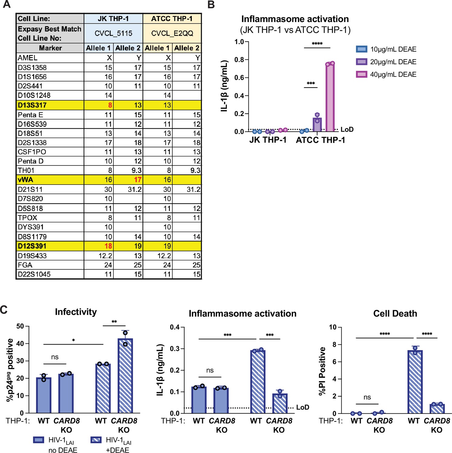
There have been four different sublines of THP-1 cells previously characterized (Kasai et al., 2022). Using short tandem repeat (STR) profiling, we were able to distinguish the WT THP-1 cell stocks that were used as the parental line for knockouts and complemented knockouts in this work and our prior work (Kulsuptrakul et al., 2023), as distinct from WT THP-1 cells sourced from ATCC at three different loci (Appendix 1—figure 1A). Of note, unlike the THP-1 cells used here (referred to as JK THP-1), ATCC THP-1 cells elicited IL-1β secretion in the absence of HIV-1 infection in the presence of 20μg/mL DEAE-dextran (Appendix 1—figure 1B). Unless otherwise specified, any mention of ‘THP-1 cells’ are referring to our JK THP-1 cells, not ATCC THP-1 cells. Nonetheless, given the sensitivity of some THP-1 sublines to elicit an inflammasome response in the presence of DEAE-dextran, we assessed whether or not we could establish systems to measure HIV-1-induced CARD8-dependent inflammasome activation in the absence of DEAE-dextran. Thus, we infected either wildtype (WT) or CARD8 knockout (KO) THP-1 cells with wildtype HIV-1LAI in either the presence or absence of DEAE-dextran and measured cell death and IL-1β secretion 24 hours post-infection as readouts of inflammasome activation. We found that despite achieving similar levels of infection (20–30%) as measured by intracellular p24gag after spinoculation with and without DEAE-dextran (Appendix 1—figure 1C, left), we detected robust CARD8-dependent inflammasome activation in WT THP-1 cells infected only in the presence of DEAE-dextran (Appendix 1—figure 1C, middle and right). These data suggest that cationic polymer is necessary to observe HIV-dependent CARD8 inflammasome activation in our cell-free system.
Appendix 1—figure 1
Characterization of THP-1 cells.
(A) Promega GenePrint 24 system STR analysis summary of our JK THP-1 cells versus ATCC THP-1 cells. Cell line authentication was done by TransnetYX, Inc by following the protocol described in ANSI/ATCC ASN-0002–2011. The STR alleles were searched on the ATCC Database and the Expasy best match cell numbers for each cell line had a 100% database match. Distinguishing loci are highlighted in yellow and distinguishing alleles are in red. (B) JK and ATCC THP-1 cells were primed with Pam3CSK4 overnight then treated with increasing doses of DEAE-dextran for 24 hours before probing for IL-1β secretion. (C) Wildtype (WT) or CARD8 knockout (KO) THP-1 cells were infected with wildtype HIV-1LAI at the same MOI in the presence or absence of DEAE-dextran (10 µg/mL) then harvested after 24 hours and assayed for: left, percent infection via intracellular p24gag; middle, inflammasome activation by IL-1β secretion via IL-1R reporter assay; and right, cell death via propidium iodide (PI) dye uptake using flow cytometry. %PI positive and IL-1 levels are normalized to mock control. Dotted line indicates limit of detection (LoD). Datasets represent mean ± SD (n=2 biological replicates). Two-way ANOVA with (B) Sidak’s or (C) Tukey’s test using GraphPad Prism 10. ns = not significant, *p<0.05, **p<0.01, ***p<0.001, ****P<0.0001.
Data availability
All data generated during this study are included in the manuscript or posted to Dryad. Sequences have been submitted to GenBank at the following accession codes (PV761644–PV761650).
-
Dryad Digital RepositoryData from: CARD8 inflammasome activation during HIV-1 cell-to-cell transmission.https://doi.org/10.5061/dryad.qbzkh18vn
-
NCBI GenBankID PV761644. Mutant HIV-1 clone CA126802 from USA gag protein (gag) and pol protein, protease region, (pol) genes, partial cds.
-
NCBI GenBankID PV761645. Mutant HIV-1 clone CA122805 from USA gag protein (gag) and pol protein, protease region, (pol) genes, partial cds.
-
NCBI GenBankID PV761646. Mutant HIV-1 clone CA96457 from USA gag protein (gag) and pol protein, protease region, (pol) genes, partial cds.
-
NCBI GenBankID PV761647. Mutant HIV-1 clone CA96458 from USA gag protein (gag) and pol protein, protease region, (pol) genes, partial cds.
-
NCBI GenBankID PV761648. Mutant HIV-1 clone CA50834-1 from USA gag protein (gag) and pol protein, protease region, (pol) genes, partial cds.
-
NCBI GenBankID PV761649. Mutant HIV-1 clone CA96451 from USA gag protein (gag) and pol protein, protease region, (pol) genes, partial cds.
-
NCBI GenBankID PV761650. Mutant HIV-1 clone CA20392-1 from USA gag protein (gag) and pol protein, protease region, (pol) genes, partial cds.
References
-
HIV cell-to-cell transmission: effects on pathogenesis and antiretroviral therapyTrends in Microbiology 23:289–295.https://doi.org/10.1016/j.tim.2015.02.003
-
Macrophage infection via selective capture of HIV-1-infected CD4+ T cellsCell Host & Microbe 16:711–721.https://doi.org/10.1016/j.chom.2014.10.010
-
Inflammasomes: mechanism of assembly, regulation and signallingNature Reviews. Immunology 16:407–420.https://doi.org/10.1038/nri.2016.58
-
Tripping the wire: sensing of viral protease activity by CARD8 and NLRP1 inflammasomesCurrent Opinion in Immunology 83:102354.https://doi.org/10.1016/j.coi.2023.102354
-
Macrophage-tropic human immunodeficiency virus isolates from different patients exhibit unusual V3 envelope sequence homogeneity in comparison with T-cell-tropic isolates: definition of critical amino acids involved in cell tropismJournal of Virology 66:6547–6554.https://doi.org/10.1128/JVI.66.11.6547-6554.1992
-
HIV-1 tat primes and activates microglial NLRP3 inflammasome-mediated neuroinflammationThe Journal of Neuroscience 37:3599–3609.https://doi.org/10.1523/JNEUROSCI.3045-16.2017
-
Chemical inhibition of DPP9 sensitizes the CARD8 inflammasome in HIV-1-infected cellsNature Chemical Biology 19:431–439.https://doi.org/10.1038/s41589-022-01182-5
-
Infection of monocyte-derived macrophages with human immunodeficiency virus type 1 (HIV-1). Monocyte-tropic and lymphocyte-tropic strains of HIV-1 show distinctive patterns of replication in a panel of cell typesThe Journal of Experimental Medicine 170:1149–1163.https://doi.org/10.1084/jem.170.4.1149
-
Inference of CRISPR edits from sanger trace dataThe CRISPR Journal 5:123–130.https://doi.org/10.1089/crispr.2021.0113
-
Multiploid inheritance of HIV-1 during cell-to-cell infectionJournal of Virology 85:7169–7176.https://doi.org/10.1128/JVI.00231-11
-
Comprehensive mapping of HIV-1 escape from a broadly neutralizing antibodyCell Host & Microbe 21:777–787.https://doi.org/10.1016/j.chom.2017.05.003
-
Peripheral blood CCR4+CCR6+ and CXCR3+CCR6+CD4+ T cells are highly permissive to HIV-1 infectionJournal of Immunology 184:1604–1616.https://doi.org/10.4049/jimmunol.0903058
-
A ubiquitin-independent proteasome pathway controls activation of the CARD8 inflammasomeThe Journal of Biological Chemistry 298:102032.https://doi.org/10.1016/j.jbc.2022.102032
-
THP-1 reference data: Proposal of an in vitro branched evolution model for cancer cell linesInternational Journal of Cancer 151:463–472.https://doi.org/10.1002/ijc.34019
-
CARD8 inflammasome activation triggers pyroptosis in human T cellsThe EMBO Journal 39:e105071.https://doi.org/10.15252/embj.2020105071
-
HIV infection stabilizes macrophage-T cell interactions to promote cell-cell HIV spreadJournal of Virology 93:e00805-19.https://doi.org/10.1128/JVI.00805-19
-
Antiretroviral Resistance in Clinical PracticeThe impact of resistance on viral fitness and its clinical implications, Antiretroviral Resistance in Clinical Practice, Mediscript.
-
HIV-1 viral protein R activates NLRP3 inflammasome in microglia: implications for HIV-1 associated neuroinflammationJournal of Neuroimmune Pharmacology 12:233–248.https://doi.org/10.1007/s11481-016-9708-3
-
Viral proteases activate the CARD8 inflammasome in the human cardiovascular systemThe Journal of Experimental Medicine 219:e20212117.https://doi.org/10.1084/jem.20212117
-
In vivo cytolysis and fusion of human immunodeficiency virus type 1-infected lymphocytes in lymphoid tissueThe Journal of Infectious Diseases 182:338–342.https://doi.org/10.1086/315640
-
Nelfinavir-resistant, amprenavir-hypersusceptible strains of human immunodeficiency virus type 1 carrying an N88S mutation in protease have reduced infectivity, reduced replication capacity, and reduced fitness and process the Gag polyprotein precursor aberrantlyJournal of Virology 76:8659–8666.https://doi.org/10.1128/jvi.76.17.8659-8666.2002
-
Prototypical recombinant multi-protease-inhibitor-resistant infectious molecular clones of human immunodeficiency virus type 1Antimicrobial Agents and Chemotherapy 57:4290–4299.https://doi.org/10.1128/AAC.00614-13
-
(S)-1-((S)-2-{[1-(4-amino-3-chloro-phenyl)-methanoyl]-amino}-3,3-dimethyl-butanoyl)-pyrrolidine-2-carboxylic acid ((2R,3S)-2-ethoxy-5-oxo-tetrahydro-furan-3-yl)-amide (VX-765), an orally available selective interleukin (IL)-converting enzyme/caspase-1 inhibitor, exhibits potent anti-inflammatory activities by inhibiting the release of IL-1beta and IL-18The Journal of Pharmacology and Experimental Therapeutics 321:509–516.https://doi.org/10.1124/jpet.106.111344
-
NLRP3 inflammasome induces CD4+ T cell loss in chronically HIV-1–infected patientsJournal of Clinical Investigation 131:38861.https://doi.org/10.1172/JCI138861
Article and author information
Author details
Jessie Kulsuptrakul

Patrick S Mitchell
Funding
National Institutes of Health (DP2 AI 154432-01)
- Patrick S Mitchell
National Institutes of Health (DP1 DA051110)
- Michael Emerman
National Institutes of Health (T32 GM007270)
- Jessie Kulsuptrakul
Howard Hughes Medical Institute
- Patrick S Mitchell
Mallinckrodt Foundation
- Patrick S Mitchell
The funders had no role in study design, data collection and interpretation, or the decision to submit the work for publication.
Acknowledgements
We thank everyone in the Emerman and Mitchell labs for helpful feedback on the project, Terry Hafer and Marisa Yonemitsu for critical reading of the manuscript, Liang Shan and his lab members for discussions and sharing of unpublished results and protocols, and the Fred Hutchinson Shared Resources Genomics, Flow Cytometry, and Specimen Processing & Research Cell Bank cores. LPV (HRP-9481), NVP (HRP-4666), and p24gag antibody (ARP-3537) were provided by the AIDS Reagent Program, Division of AIDS, NIAID, and NIH. PSM is an HHMI Freeman Hrabowski Scholar and is supported by grants from the National Institutes of Health (NIH) (DP2 AI 154432-01) and the Mallinckrodt Foundation to PSM. ME is supported by NIH grant DP1 DA051110. JK is supported by the University of Washington Cellular and Molecular Biology Training Grant (T32 GM007270).
Version history
- Preprint posted:
- Sent for peer review:
- Reviewed Preprint version 1:
- Reviewed Preprint version 2:
- Version of Record published:
Cite all versions
You can cite all versions using the DOI https://doi.org/10.7554/eLife.102676. This DOI represents all versions, and will always resolve to the latest one.
Copyright
© 2024, Kulsuptrakul et al.
This article is distributed under the terms of the Creative Commons Attribution License, which permits unrestricted use and redistribution provided that the original author and source are credited.
Metrics
-
- 1,450
- views
-
- 125
- downloads
-
- 5
- citations
Views, downloads and citations are aggregated across all versions of this paper published by eLife.
Citations by DOI
-
- 1
- citation for umbrella DOI https://doi.org/10.7554/eLife.102676
-
- 4
- citations for Version of Record https://doi.org/10.7554/eLife.102676.3
Download links
A two-part list of links to download the article, or parts of the article, in various formats.
Downloads (link to download the article as PDF)
Open citations (links to open the citations from this article in various online reference manager services)
Cite this article (links to download the citations from this article in formats compatible with various reference manager tools)
CARD8 inflammasome activation during HIV-1 cell-to-cell transmission
eLife 13:RP102676.
https://doi.org/10.7554/eLife.102676.3


