Neuronal temperature perception induces specific defenses that enable C. elegans to cope with the enhanced reactivity of hydrogen peroxide at high temperature

  1. Francesco A Servello
  2. Rute Fernandes
  3. Matthias Eder
  4. Nathan Harris
  5. Olivier MF Martin
  6. Natasha Oswal
  7. Anders Lindberg
  8. Nohelly Derosiers
  9. Piali Sengupta
  10. Nicholas Stroustrup
  11. Javier Apfeld  Is a corresponding author
  1. Biology Department, Northeastern University, United States
  2. Centre for Genomic Regulation (CRG), The Barcelona Institute of Science and Technology, Spain
  3. Universitat Pompeu Fabra (UPF), Spain
  4. Department of Biology, Brandeis University, United States
  5. Bioengineering Department, Northeastern University, United States

Abstract

Hydrogen peroxide is the most common reactive chemical that organisms face on the microbial battlefield. The rate with which hydrogen peroxide damages biomolecules required for life increases with temperature, yet little is known about how organisms cope with this temperature-dependent threat. Here, we show that Caenorhabditis elegans nematodes use temperature information perceived by sensory neurons to cope with the temperature-dependent threat of hydrogen peroxide produced by the pathogenic bacterium Enterococcus faecium. These nematodes preemptively induce the expression of specific hydrogen peroxide defenses in response to perception of high temperature by a pair of sensory neurons. These neurons communicate temperature information to target tissues expressing those defenses via an insulin/IGF1 hormone. This is the first example of a multicellular organism inducing their defenses to a chemical when they sense an inherent enhancer of the reactivity of that chemical.

Editor's evaluation

For any organism, tailoring defenses to the most pressing threats has high adaptive value – this paper makes the important finding that the nematode C. elegans pre-emptively augments its defenses against hydrogen peroxide when temperature increases, a condition that enhances the damage this compound causes. The authors describe this strategy as ‘enhancer sensing,’ whereby the perception of an environmental stimulus leads to the induction of defenses against a distinct (but mechanistically linked) threat. Convincing mechanistic studies reveal a role for a key thermosensory neuron and insulin-like signaling in this phenomenon. Because of its interdisciplinary outlook, this work will be of interest to readers in the fields of sensory neuroscience, stress physiology, and evolutionary biology.

https://doi.org/10.7554/eLife.78941.sa0

eLife digest

The Earth’s environment is full of reactive chemicals that can cause harm to organisms. One of the most common is hydrogen peroxide, which is produced by several bacteria in concentrations high enough to kill small animals, such as the roundworm Caenorhabditis elegans. Forced to live in close proximity to such perils, C. elegans have evolved defenses to ensure their survival, such as producing enzymes that can break down hydrogen peroxide.

However, this battle is compounded by other factors. For instance, rising temperatures can increase the rate at which the hydrogen peroxide produced by bacteria reacts with the molecules and proteins of C. elegans. In 2020, a group of researchers found that roundworms sense these temperature changes through special cells called sensory neurons and use this information to control the generation of enzymes that break down hydrogen peroxide. This suggests that C. elegans may pre-emptively prepare their defenses against hydrogen peroxide in response to higher temperatures so they are better equipped to shield themselves from this harmful chemical.

To test this theory, Servello et al. – including some of the authors involved in the 2020 study – exposed C. elegans to a species of bacteria that produces hydrogen peroxide. This revealed that the roundworms were better at dealing with the threat of hydrogen peroxide when growing in warmer temperatures. Experiments done in C. elegans lacking a class of sensory cells, the AFD neurons, showed that these neurons increased the roundworms’ resistance to the chemical when temperatures increase. They do this by repressing the activity of INS-39, a hormone that stops C. elegans from switching on their defense mechanism against peroxides.

This is the first example of a multicellular organism preparing its defenses to a chemical after sensing something (such as temperature) that enhances its reactivity. It is possible that other animals may also use this ‘enhancer sensing' strategy to anticipate and shield themselves from hydrogen peroxide and potentially other external threats.

Introduction

Reactive chemicals in the environment pose a lethal threat to organisms by changing the chemical composition of their macromolecules. But organisms are not passive chemical substrates, they have sophisticated defense mechanisms that deal with the threat posed by those chemicals. That threat is inherently temperature dependent because chemical reactions occur at faster rates at higher temperatures (Arrhenius, 1889; Evans and Polanyi, 1935; Eyring, 1935). However, the extent to which the defense mechanisms protecting the organism from reactive chemicals are adjusted to balance the temperature-dependent threat posed by those chemicals remains poorly understood. In the present study, we used the nematode Caenorhabditis elegans as a model system to explore the extent to which temperature regulates how multicellular organisms deal with the threat of hydrogen peroxide.

Hydrogen peroxide (H2O2) is the most common reactive chemical that organisms face on the microbial battlefield (Mishra and Imlay, 2012). Bacteria, fungi, plants, and animal cells have long been known to use H2O2 as an offensive weapon that damages the nucleic acids, proteins, and lipids of their targets (Avery and Morgan, 1924; Imlay, 2018). C. elegans encounter a wide variety of bacteria in their ecological setting (Samuel et al., 2016; Schiffer et al., 2021), including many genera known to produce H2O2 (Passardi et al., 2007). H2O2 produced by a bacterium from the C. elegans microbiome, Neorhizobium sp., causes DNA damage to the nematodes (Kniazeva and Ruvkun, 2019) and many bacteria—including Enterococcus faecium, Streptococcus pyogenes, Streptococcus pneumoniae, and Streptococcus oralis—kill C. elegans by producing millimolar concentrations of hydrogen peroxide (Bolm et al., 2004; Jansen et al., 2002; Moy et al., 2004).

Prevention and repair of the damage that hydrogen peroxide inflicts on macromolecules are critical for cellular health and survival (Chance et al., 1979). To avoid damage from H2O2, C. elegans rely on conserved cellular defenses, including H2O2-degrading catalases (Chávez et al., 2007; Schiffer et al., 2020). However, inducing those defenses at inappropriate times can cause undesirable side effects, including developmental defects (Doonan et al., 2008; Kramer-Drauberg et al., 2020), because H2O2 modulates the function of proteins involved in a wide variety of cellular processes, including signal transduction and differentiation (Hourihan et al., 2016; Kramer-Drauberg et al., 2020; Meng et al., 2021; Veal et al., 2007). We recently found that 10 classes of sensory neurons in the brain of C. elegans manage the challenge of deciding when the nematode’s tissues induce H2O2 defenses (Schiffer et al., 2020). Sensory neurons might be able to integrate a wider variety of inputs than the individual tissues expressing those defenses could integrate, enabling a better assessment of the threat of hydrogen peroxide.

In their habitat, C. elegans face daily and seasonal variations in temperature, which can affect a wide variety of processes, including development, reproduction, and lifespan (Golden and Riddle, 1984; Klass, 1977). Temperature also affects the growth of bacteria (Barber, 1908; Rosso et al., 1993) and, therefore, likely affects the interactions of C. elegans with beneficial and pathogenic bacteria (Samuel et al., 2016; Zhang et al., 2017). While nematodes do not regulate their own body temperature, they adjust their behavior and physiology in response to the perception of temperature by sensory neurons, enabling them to seek temperatures conducive to survival, avoid noxious temperature ranges, and induce heat defenses (Goodman and Sengupta, 2019; Hedgecock and Russell, 1975; Prahlad et al., 2008). Nematodes perceive temperature, in part, via seven classes of sensory neurons (Beverly et al., 2011; Biron et al., 2008; Chatzigeorgiou et al., 2010; Kuhara et al., 2008; Liu et al., 2012; Mori and Ohshima, 1995; Schild et al., 2014). Previously, we found that four of those classes of neurons regulate C. elegans peroxide resistance (Schiffer et al., 2020), suggesting that nematodes might adjust their peroxide defenses in response to temperature perception.

Here, we show that the lethality of hydrogen peroxide to C. elegans increases with temperature. Nematodes partially compensate for this by preemptively inducing their hydrogen peroxide defenses at high temperature. This adaptive response to temperature enables the nematodes to better cope with H2O2 produced by the pathogenic bacterium E. faecium. The temperature-dependent regulation of peroxide defenses is directed by the AFD sensory neurons. At high temperature, the AFD neurons repress the expression of the INS-39 insulin/IGF1 hormone and thereby alleviate inhibition by insulin/IGF1 signaling of the nematodes’ peroxide defenses. The insulin/IGF1 effector DAF-16/FOXO functions in intestinal cells to determine the size of the gene-expression changes induced by the absence of signals from the AFD neurons. By coupling the induction of H2O2 defenses to the perception of high temperature—an inherent enhancer of the reactivity of H2O2—the nematodes are assessing faithfully the threat that H2O2 poses.

Results

C. elegans induces long-lasting peroxide defenses in response to high temperature

In their natural habitat, C. elegans nematodes encounter many threats that can shorten their lifespan. A major chemical threat that C. elegans face is hydrogen peroxide (H2O2). Bacteria can produce millimolar concentrations of H2O2 (Bolm et al., 2004; Jansen et al., 2002; Moy et al., 2004), shortening C. elegans lifespan over tenfold (Chávez et al., 2007). A major physical threat that C. elegans face is high temperature. Even a small increase in temperature from 20 to 25°C—within the range of ambient temperatures that C. elegans prefers in nature (Crombie et al., 2019)—shortens C. elegans lifespan from 19 to 15 days (Klass, 1977; Lee and Kenyon, 2009; Stroustrup et al., 2016). We set out to investigate the extent to which H2O2 and temperature acted together to determine C. elegans survival.

Previous studies showed that H2O2 kills C. elegans in a dose-dependent manner at environmental concentrations above 0.1 mM (Bolm et al., 2004; Jansen et al., 2002; Moy et al., 2004). We expected that higher temperatures would make the same concentration of H2O2 more lethal to C. elegans, because the reaction rates of the chemical reactions of H2O2 increase exponentially with temperature (Arrhenius, 1889; Evans and Polanyi, 1935; Eyring, 1935). The exact molecular mechanisms by which H2O2 kills C. elegans, or any organism, remain unknown but are thought to involve the reactions of H2O2 with biologically important molecules, including proteins and DNA (Khademian and Imlay, 2021). Using chemical kinetics, we modeled how an increase in temperature from 20 to 25°C would affect the rates of the chemical reactions of H2O2 with those biomolecules (Figure 1). Because these rate differences depend on the enthalpy of activation of the specific chemical reaction, they can vary widely between reactions. The Fenton reaction of H2O2 with DNA-bound Fe(II), which leads to DNA damage, was predicted to be 40% faster at 25°C than at 20°C (Figure 1). For the oxidation of the thiol groups of cysteines, reaction rates with H2O2 were predicted to be more than twofold faster for regular cysteines in proteins, 62% faster for free cysteines, up to 56% faster for very reactive cysteines such as the redox-sensitive cysteine residue of GAPDH, and 17% faster for the most reactive cysteines of hydroperoxidases (Figure 1). These predicted increases in H2O2’s reactivity toward specific biomolecules at 25°C, compared to 20°C, are similar to the ones that would occur at 20°C if H2O2 concentration were increased substantially—from 17% to more than 100%, depending on the specific reaction.

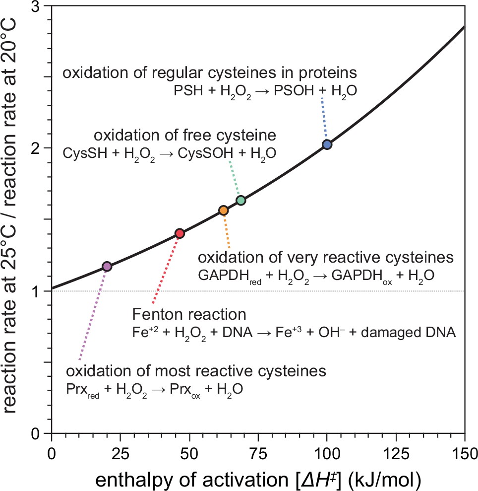
Temperature-dependent enhancement of H2O2 reactivity toward biologically important molecules.

Using chemical kinetics, we modeled how much the rates of chemical reactions of H2O2 change between 20 and 25°C. This plot illustrates how the enthalpy of activation of a specific chemical reaction influences its rate at 25°C compared to 20°C. The dotted arrows point to the relative reaction rate values for specific reactions of H2O2 with biologically important molecules.

To investigate the extent to which cultivation temperature might influence C. elegans survival in the presence of environmental peroxides, we measured the peroxide resistance of nematodes cultured within their preferred temperature range (Crombie et al., 2019). We cultured the nematodes at either 20 or 25°C until the second day of adulthood, and then determined their subsequent survival at those temperatures in the presence of a peroxide in their environment. We used tert-butyl hydroperoxide (tBuOOH) because this peroxide, unlike H2O2, is not degraded efficiently by Escherichia coli—the nematodes’ conventional food in the laboratory (Brenner, 1974). Previously, we found that when tBuOOH concentration exceeded 0.75 mM, C. elegans lifespan was shortened by 50% by each additional 45% increase in tBuOOH concentration (Stroustrup et al., 2016). In the presence of 6 mM tBuOOH, nematodes grown at 20°C survived an average of 1.6 days at 20°C, while those grown and assayed at 25°C survived 30% shorter (Figure 2A). Therefore, C. elegans peroxide resistance was temperature dependent.

Figure 2 with 1 supplement see all
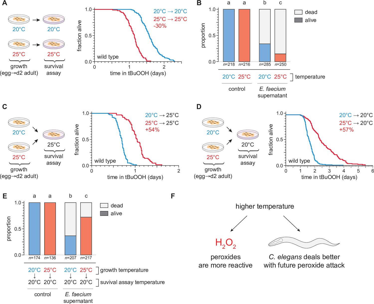
Temperature regulates the peroxide resistance of C. elegans.

(A) Peroxide resistance of wild-type C. elegans grown and assayed at 20 or 25°C. The fraction of nematodes remaining alive in the presence of 6 mM tert-butyl hydroperoxide (tBuOOH) is plotted against time. (B) Survival of wild-type C. elegans 16 hr after exposure to E. faecium E007 liquid-culture supernatant. Nematodes were grown and assayed at 20 or 25°C. Groups labeled with different letters exhibited significant survival differences (p < 0.001, ordinal logistic regression) otherwise (p > 0.05). (C) Peroxide resistance at 25°C of wild-type C. elegans grown at 20 or 25°C. (D) Peroxide resistance at 20°C of wild-type C. elegans grown at 20 or 25°C. (E) Survival of wild-type C. elegans 16 hr after exposure to E. faecium E007 liquid-culture supernatant. Nematodes were grown at 20 or 25°C and assayed at 20°C. Groups labeled with different letters exhibited significant survival differences (p < 0.001, ordinal logistic regression) otherwise (p > 0.05). (F) Peroxides killed C. elegans more quickly at 25°C than at 20°C, but nematodes grown at 25°C could survive a subsequent peroxide exposure better than those grown at 20°C. Statistical analyses for panels (A, C, and D) are in Supplementary file 1.

We speculated that C. elegans survival to bacterially produced H2O2 would, likewise, be shorter at 25°C than at 20°C. H2O2 produced by the pathogenic bacterium E. faecium is lethal to C. elegans (Chávez et al., 2007; Moy et al., 2004). We exposed day 2 adult nematodes that fed on E. coli JI377—a katG katE ahpCF triple null mutant strain which cannot degrade environmental H2O2 (Seaver and Imlay, 2001)—to the supernatant of an E. faecium liquid culture and, after 16 hr, determined the proportion of nematodes that survived. Compared to nematodes grown and assayed at 20°C, those grown and assayed at 25°C were less likely to survive the E. faecium supernatant (Figure 2B), indicating that H2O2 was more lethal to C. elegans at the higher temperature. Together, these observations indicated that at the upper end of the natural temperature range of C. elegans, two types of peroxides were more lethal to the nematodes.

We expected that increasing temperature would make peroxides more lethal to C. elegans because temperature increases the rate of chemical reactions, including those that mediate peroxide-dependent killing. If this was the only mechanism by which temperature affected C. elegans’ peroxide resistance, then peroxide resistance should have been determined by the temperature the nematodes experienced during the peroxide resistance assay and not by the temperature they experienced before they were exposed to peroxide. Alternatively, the temperature C. elegans experienced before encountering peroxides in the environment may have influenced their subsequent sensitivity to peroxide. For example, a high cultivation temperature may have irreversibly damaged the nematodes, thus rendering them more sensitive to peroxide-dependent killing.

To distinguish between these possibilities, we measured the effects of the nematodes’ growth-temperature history (before peroxide exposure) on their subsequent peroxide resistance by performing temperature-shift experiments where nematode populations grown at 20 or 25°C were transferred to assay plates containing 6 mM tBuOOH at either 20 or 25°C. To our surprise, we found that in survival assays performed at 25°C the nematodes grown at 25°C lived 54% longer than those grown at 20°C (Figure 2C). Similarly, in assays performed at 20°C, the nematodes grown at 25°C lived 57% longer than those grown at 20°C (Figure 2D). We also found that, compared with nematodes grown at 20°C, a higher proportion of nematodes grown at 25°C survived exposure to E. faecium liquid-culture supernatant at 20°C (Figure 2E). Therefore, nematodes grown at 25°C were more peroxide resistant than those grown at 20°C.

Our findings contradicted a model where temperature affected how quickly the nematodes were killed by peroxides only by influencing the reactivity of peroxides. In addition, those findings contradicted a prediction that high temperature would irreversibly render the nematodes more sensitive to peroxide-dependent killing. Instead, we conclude that even though peroxides killed C. elegans more quickly at 25°C than at 20°C, nematodes grown at 25°C could better survive a subsequent peroxide exposure than those grown at 20°C. Based on these findings, we speculated that C. elegans nematodes induced their peroxide defenses when grown at the higher temperature to prepare for the increased lethal threat posed by peroxides at high temperature (Figure 2F).

To determine the extent to which these differences in the nematodes’ growth temperature had lasting effects on their subsequent peroxide resistance, we repeated the temperature-shift experiments, but this time we transferred the nematodes to the higher or lower temperature 1 day before the peroxide survival assay (on day 1 of adulthood), and 2 days before (at the onset of adulthood). Shifting from 20 to 25°C for 2 days was sufficient to improve peroxide survival at 25°C, but shifting only 1 day before the assay was not sufficient (Figure 2—figure supplement 1A). Therefore, nematodes grown at 20°C could increase their peroxide resistance in response to a temperature increase during adulthood. Nematodes down-shifted from 25 to 20°C for 2 days, 1 day, or immediately before the assay were all more peroxide resistant at 20°C than those grown continuously at 20°C (Figure 2—figure supplement 1B). Therefore, growth at 25°C could increase the nematodes’ peroxide resistance even days after they had been transferred to 20°C. Together, these observations suggested that C. elegans can slowly induce long-lasting peroxide defenses in response to the higher cultivation temperature.

AFD sensory neurons are required for the temperature dependence of C. elegans peroxide resistance

We recently found that sensory neurons regulate C. elegans sensitivity to peroxides in the environment (Schiffer et al., 2020). To investigate whether temperature might regulate C. elegans peroxide defenses via sensory neurons, we determined whether mutations that cause defects in the transduction of sensory information within neurons affected the extent to which temperature influenced the nematodes’ peroxide resistance. We examined tax-4 cyclic GMP-gated channel mutants, which are defective in the transduction of several sensory stimuli, including temperature (Coburn and Bargmann, 1996; Komatsu et al., 1996). When grown at 20°C, tax-4 mutants exhibited an over twofold increase in peroxide resistance at 20°C relative to wild-type controls (Figure 3A, B). In contrast, when grown and assayed at 25°C, tax-4 mutants exhibited a smaller increase in peroxide resistance, 49% (Figure 3C). These findings suggested that neuronal sensory transduction by TAX-4 channels normally lowers C. elegans’ peroxide resistance to a lesser extent at high cultivation temperature.

Figure 3 with 1 supplement see all
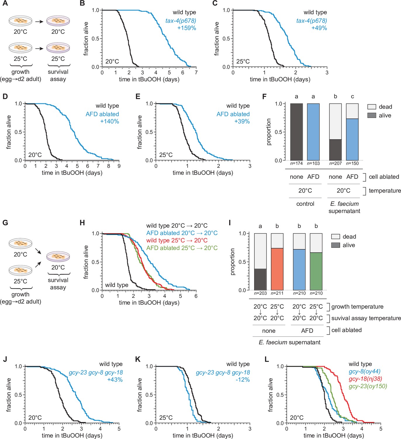
The AFD sensory neurons are required for the temperature dependence of C.elegans peroxide resistance.

(A) Diagram summarizing the experimental strategy for panels (B–F and J–L). The tax-4(p678) mutation increased peroxide resistance by a greater factor at 20°C (B) than at 25°C (C). Genetic ablation of AFD increased peroxide resistance by a greater factor at 20°C (D) than at 25°C (E). (F) Genetic ablation of AFD increased the proportion of nematodes that survived 16 hr after exposure to E. faecium E007 liquid-culture supernatant. Nematodes were grown and assayed at 20°C. Groups labeled with different letters exhibited significant differences (p < 0.001, ordinal logistic regression) otherwise (p > 0.05). (G) Diagram summarizing the experimental strategy for panels (H, I). (H) AFD-ablated nematodes grown at 25°C did not exhibit a further increase in peroxide resistance at 20°C, unlike wild-type (unablated) nematodes. (I) Growth at 25°C did not further increase the proportion of AFD-ablated nematodes that survived after 16-hr exposure to E. faecium E007 liquid-culture supernatant at 20°C, unlike in wild-type (unablated) nematodes. Groups labeled with different letters exhibited significant differences (p < 0.001, ordinal logistic regression) otherwise (p > 0.05). (J–K) gcy-23(oy150) gcy-8(oy44) gcy-18(nj38) triple null mutants show increased peroxide resistance at 20°C (J) and decreased peroxide resistance at 25°C (J). (L) Peroxide resistance of gcy-8(oy44), gcy-18(nj38), and gcy-23(oy150) single mutants and wild-type nematodes at 20°C. Statistical analyses for panels (B–E, H, J–L) are in Supplementary file 2.

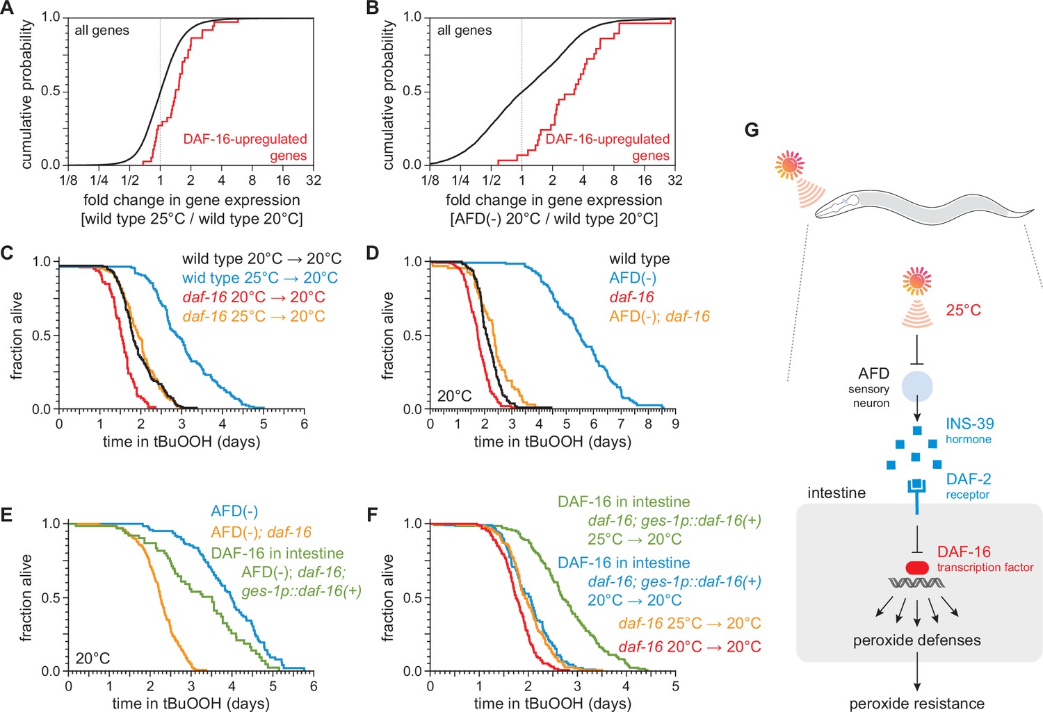
To identify specific neurons that regulate C. elegans peroxide defenses in response to temperature, we focused on a single pair of neurons, the AFD neurons, chosen from the small subset of sensory neurons in which TAX-4 channels are expressed (Coburn and Bargmann, 1996; Komatsu et al., 1996). Previously, we found that genetic ablation of the AFD neurons via neuron-specific expression of caspase (Chelur and Chalfie, 2007; Glauser et al., 2011) increased C. elegans peroxide resistance (Schiffer et al., 2020). Because the AFD neurons respond to temperature via TAX-4 channels to regulate diverse temperature-dependent behaviors (Hedgecock and Russell, 1975; Mori and Ohshima, 1995), we speculated that they might also regulate peroxide resistance in response to temperature. To determine whether AFD neurons lowered the nematodes’ peroxide resistance in a temperature-dependent manner, we measured the effects of AFD ablation at 20 and 25°C. Compared with wild-type nematodes, when grown and assayed at 20°C, the AFD-ablated nematodes exhibited an over twofold increase in resistance to tBuOOH (Figure 3D) and H2O2 (Figure 3—figure supplement 1), and were more likely to survive exposure to E. faecium liquid-culture supernatant (Figure 3F). At 25°C, AFD ablation increased resistance to tBuOOH by 31% (Figure 3E), a much smaller amount than at 20°C. Therefore, the AFD neurons normally lower C. elegans peroxide resistance in a temperature-dependent manner.

If the AFD neurons were blocking the induction of peroxide defenses, we hypothesized that ablation of both AFD neurons might result in induction of peroxide defenses at lower temperatures similar to those seen in unablated nematodes at the higher temperature. Therefore, we predicted that AFD-ablated nematodes grown at 25°C would not exhibit a further increase in peroxide resistance at 20°C, unlike wild-type nematodes. Consistent with that prediction, AFD-ablated nematodes grown at 25°C exhibited the same levels of resistance as wild-type nematodes grown at 25°C (Figure 3G, H). AFD-ablated nematodes grown continuously at 20°C exhibited the highest levels of peroxide resistance (Figure 3H). Similarly, in assays at 20°C measuring nematode survival after exposure to a supernatant derived from a liquid culture of E. faecium, AFD-ablated nematodes grown at either 20 or 25°C survived as well as wild-type nematodes grown at 25°C (Figure 3I). We propose that, in wild-type C. elegans, the extent to which the AFD neurons lower peroxide defenses is reduced in response to higher temperature.

Last, we determined whether previously identified mechanisms for temperature perception by the AFD neurons were required for the temperature-dependent regulation of peroxide resistance. The AFD neurons sense temperature using receptor guanylate cyclases, which catalyze cGMP production, leading to the opening of TAX-4 channels (Goodman and Sengupta, 2019). Three receptor guanylate cyclases are expressed exclusively in AFD neurons: GCY-8, GCY-18, and GCY-23 (Inada et al., 2006; Yu et al., 1997) and are thought to act as temperature sensors (Takeishi et al., 2016). Triple mutants lacking gcy-8, gcy-18, and gcy-23 function are behaviorally atactic on thermal gradients and fail to display changes in intracellular calcium or thermoreceptor current in the AFD neurons in response to temperature changes (Inada et al., 2006; Ramot et al., 2008; Takeishi et al., 2016; Wang et al., 2013; Wasserman et al., 2011). We found that when grown and assayed at 20°C, gcy-23(oy150) gcy-8(oy44) gcy-18(nj38) triple null mutants survived 43% longer in the presence of tBuOOH than wild-type controls (Figure 3J). In contrast, at 25°C, the gcy-23 gcy-8 gcy-18 triple mutants showed a 12% decrease in peroxide resistance relative to wild-type controls (Figure 3K). Therefore, the three AFD-specific receptor guanylate cyclases influenced the temperature dependence of peroxide resistance, lowering peroxide resistance at 20°C and slightly increasing it at 25°C. At 20°C, the gcy-8(oy44), gcy-18(nj38), and gcy-23(oy150) single mutants increased peroxide resistance by 10%, 51%, and 21%, respectively, relative to wild-type controls (Figure 3L). Therefore, each of the three AFD-specific receptor guanylate cyclases regulates peroxide resistance, and their roles are not fully redundant. We conclude that temperature perception by AFD via GCY-8, GCY-18, and GCY-23 enables C. elegans to lower their peroxide resistance at the lower cultivation temperature. Other mechanisms within AFD likely contribute to the regulation of peroxide resistance, as AFD ablation caused a greater increase in peroxide resistance than the gcy-23 gcy-8 gcy-18 triple mutant.

Hydrogen peroxide defenses are induced by high cultivation temperature and by AFD ablation

To investigate whether the higher cultivation temperature and the ablation of the AFD sensory neurons increased C. elegans peroxide resistance through a common defense mechanism, we used mRNA sequencing (mRNA-seq) to compare the extent to which those interventions affected gene expression. Collecting mRNA from day 2 adults grown at 20 and 25°C and from AFD-ablated and unablated (wild-type) nematodes grown at 20°C, we identified differentially expressed transcripts. Relative to nematodes grown at 20°C, those grown at 25°C had lower expression of 2446 genes and higher expression of 809 genes, out of 18039 genes detected (q value <0.01) (Figure 4A and Figure 4—figure supplement 1A). These changes in gene expression were consistent with previous studies comparing gene expression in nematodes grown at 20 and 25°C (Gómez-Orte et al., 2018; Figure 4—figure supplement 2A, B) and in nematodes shifted from 23 to 17°C (Sugi et al., 2011; Figure 4—figure supplement 2D, E). AFD ablation lowered the expression of 2077 genes and increased the expression of 2225 genes, out of 7912 genes detected (q value <0.01) (Figure 4B and Figure 4—figure supplement 1B). Therefore, both the higher cultivation temperature and the ablation of the AFD sensory neurons induced broad changes in gene expression.

Figure 4 with 5 supplements see all
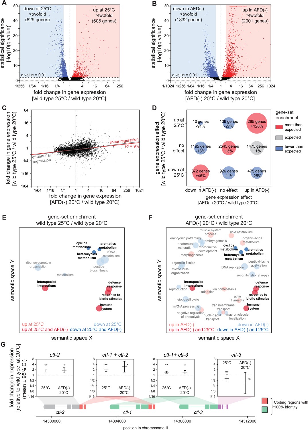
Hydrogen peroxide defenses are induced by high cultivation temperature and AFD ablation.

Volcano plots showing the level and statistical significance of changes in gene expression induced (A) in wild-type nematodes by growth at 25°C relative to growth at 20°C and (B) by AFD ablation in nematodes grown at 20°C relative to wild-type (unablated) nematodes grown at 20°C. Genes up- and downregulated significantly (q value <0.01) are shown in red and blue, respectively. (C) Growth at 25°C and AFD ablation at 20°C induced correlated changes in gene expression. Linear regression fit is shown as a red line flanked by a red area marking the 95% confidence interval of the fit. The orthogonal regression fit (gray line) makes no assumptions about the dependence or independence of the variables. (D) Coregulation of genes up- and downregulated significantly (q value <0.01) by growth at 25°C and by AFD ablation at 20°C. Bubble size is proportional to gene-set enrichment (observed/expected). Gene sets with significantly more or fewer genes than expected (p < 0.001, cell chi-square test) are colored red and blue, respectively; gene sets of the expected size (p > 0.05) are colored gray. (E, F) Gene Ontology (GO) term enrichment analysis. (E) Biological processes associated with the set of 508 upregulated genes (red bubbles) and the set of 629 downregulated genes (blue bubbles) with a statistically significant and greater than twofold change in expression in wild-type nematodes grown at 25°C relative to those grown at 20°C. (F) Biological processes associated with the set of 2001 upregulated genes (red bubbles) and the set of 1832 downregulated genes (blue bubbles) with a statistically significant and greater than twofold change in expression in AFD-ablated nematodes grown at 20°C relative to wild-type (unablated) nematodes grown at 20°C. Bubble size is proportional to the statistical significance [−log10(p value)] of enrichment. Biological processes that were induced or repressed by both interventions are bolded and shaded with darker red and blue colors, respectively. (G) Average changes in expression and 95% confidence intervals induced by growth at 25°C and AFD ablation at 20°C within intervals in the genomic region encoding the three C. elegans catalase genes. Gene models show the positions and splicing pattern of each catalase gene, intervals with 100% nucleotide identity are shown in orange (ctl-1 and ctl-2) and green (ctl-1 and ctl-3), and unique intervals are show in gray (ctl-2) and purple (ctl-3). The asterisks mark gene regions with significant fold-change in expression: **p < 0.001 and *p < 0.025, otherwise ‘ns’ indicates p > 0.1 (generalized linear model).

Figure 4—source data 1

mRNA sequencing (mRNA-seq) analysis data.

https://cdn.elifesciences.org/articles/78941/elife-78941-fig4-data1-v2.xlsx
Figure 4—source data 2

mRNA sequencing (mRNA-seq) analysis data for the genomic region of the three catalase genes.

https://cdn.elifesciences.org/articles/78941/elife-78941-fig4-data2-v2.xlsx

Next, we asked whether higher cultivation temperature and ablation of the AFD neurons altered gene expression for each transcript by the same amount and in the same direction. We found that both interventions induced changes in gene expression that were linearly correlated in a positive manner (R2 = 9%, p < 0.0001, Figure 4C). We then asked whether this weak correlation was due to co-induction of upregulated genes, co-repression of downregulated genes, or both, using categorical analysis. We found that genes with either higher or lower expression in both wild-type nematodes at 25°C and AFD-ablated nematodes at 20°C were disproportionally enriched, and that almost no genes were upregulated at 25°C but downregulated in AFD-ablated nematodes at 20°C (Figure 4D). Therefore, we conclude that cultivation temperature and AFD ablation induced overlapping changes in gene expression.

We then determined whether genes previously shown to be regulated between various temperature ranges were co-regulated by growth at 25°C and by ablation of AFD at 20°C. Genes expressed at a higher level at 25°C than at 20°C (Gómez-Orte et al., 2018) were upregulated by ablation of AFD at 20°C and were also, as expected, upregulated by growth at 25°C (Figure 4—figure supplement 2A); however, genes expressed at a higher level at 15°C than at 20°C (Gómez-Orte et al., 2018) were downregulated by growth at 25°C but were upregulated by ablation of AFD at 20°C (Figure 4—figure supplement 2B). In addition, genes induced more than twofold when nematodes at 25°C were heat shocked by shifting them to 30°C (McCarroll et al., 2004) were upregulated by ablation of AFD at 20°C, but were unaffected by growth at 25°C (Figure 4—figure supplement 2C). We conclude that, in nematodes cultivated at 20°C, the AFD sensory neurons not only repressed genes induced at a higher cultivation temperature (25°C), but also repressed genes induced at a lower cultivation temperature (15°C) and in response to heat shock (30°C).

To identify processes that may be influenced by the transcriptomic changes induced by the higher cultivation temperature and by the ablation of the AFD neurons, we used Gene Ontology (GO) term enrichment analysis (Angeles-Albores et al., 2016; Ashburner et al., 2000) and clustered enriched GO terms based on semantic similarity (Supek et al., 2011), focusing on genes with more than a twofold increase or decrease in expression between wild-type nematodes at 25 and 20°C and between AFD-ablated and unablated nematodes at 20°C. We found that both higher emperature and AFD ablation downregulated genes associated with reproduction and with expression in the germline, and upregulated genes associated with defense and immune responses and with expression in the intestine (Figure 4E, F and Supplementary file 4). To expand this analysis, we determined the extent to which higher cultivation temperature and ablation of AFD co-regulated the expression of gene sets affecting similar biological processes. We assigned each gene to a set of nested categories based on their physiological function and then their molecular function or cellular location using WormCat annotations (Higgins et al., 2022; Holdorf et al., 2020). Higher temperature and AFD ablation induced positively correlated changes in the average expression of those gene sets (R2 = 24%, p < 0.0001, Figure 4—figure supplement 3). Therefore, the higher cultivation temperature and ablation of the AFD sensory neurons appeared to induce consistent changes in the expression of genes affecting similar biological processes.

We next determined whether genes induced when nematodes were exposed to tert-butyl hydroperoxide (Oliveira et al., 2009) were also induced by the higher cultivation temperature and by the ablation of the AFD neurons. Both growth at 25°C and ablation of AFD at 20°C increased the expression of those genes (Figure 4—figure supplement 4). Therefore, in the absence of peroxide exposure, genes induced by peroxides were pre-induced in both nematodes cultivated at the higher temperature and in AFD-ablated nematodes, suggesting those nematodes were better prepared to deal with peroxides in the environment.

To identify specific peroxide defenses induced by the higher cultivation temperature and by the ablation of the AFD neurons, we focused on the catalase genes, which encode enzymes that degrade hydrogen peroxide (Loew, 1901; Nicholls, 2012; Togo et al., 2000). The C. elegans genome contains three catalase genes in tandem—two newly duplicated cytosolic catalases, ctl-1 and ctl-3, and a peroxisomal catalase, ctl-2 (Petriv and Rachubinski, 2004)—that when overexpressed 10-fold increase C. elegans resistance to hydrogen peroxide 2.7-fold (Schiffer et al., 2020). ctl-1 and ctl-2 can increase C. elegans resistance to H2O2-dependent killing (Chávez et al., 2007; Schiffer et al., 2020). Previously, we found that ctl-1 mRNA levels were 69% higher in daf-1 Type 1 TGFβ receptor loss-of-function mutants, and that ctl-1 function was required for a large part of the more than doubling of H2O2 resistance induced by those mutants (Schiffer et al., 2020). In our mRNA-seq analysis, wild-type nematodes grown at 25°C had 46% higher levels of ctl-1 expression and 73% higher levels of ctl-2 expression compared to nematodes grown at 20°C (Figure 4G), and ablation of the AFD neurons increased ctl-1 expression by 46% and increased ctl-2 expression by 89% (Figure 4G). Therefore, the cultivation temperature and the AFD neurons regulated the expression of hydrogen peroxide defenses.

Last, we determined the extent to which the higher cultivation temperature and the ablation of the AFD neurons affected the expression of genes induced by toxic organic compounds, toxic metals, and radiation (Eom et al., 2014; Greiss et al., 2008; Huffman et al., 2004; Lewis et al., 2009; Mueller et al., 2014; Sahu et al., 2013; Starnes et al., 2016). Growth at 25°C did not increase the expression of genes induced by acrylamide, formaldehyde, benzene, silver, cadmium, arsenic, UVB rays, X rays, and gamma rays (Figure 4—figure supplement 5A–I), but ablation of AFD at 20°C induced all of those gene sets (Figure 4—figure supplement 5A–I). Therefore, ablation of the AFD sensory neurons induced genes normally induced by a wide variety of stressors in nematodes that were not exposed to those stressors, but the higher cultivation temperature only pre-induced a specific subset of genes that included hydrogen peroxide defenses and genes induced by peroxides.

The high temperature-repressed INS-39 insulin/IGF1 hormone from the AFD sensory neurons lowers the nematode’s peroxide resistance

To investigate how the AFD sensory neurons regulated the nematode’s peroxide resistance, we took a candidate gene approach. We speculated that the AFD neurons signaled to target tissues via insulin/IGF1 peptide hormones because previous studies, including our own, showed that insulin/IGF1 signaling is a major determinant of peroxide resistance in C. elegans (Schiffer et al., 2020; Tullet et al., 2008). A recent single-neuron mRNA-seq study by the C. elegans Neuronal Gene Expression Map and Network consortium (CeNGEN) showed that AFD expresses many classes of peptide-hormone coding genes, including a subset of the 40 insulin/IGF1 genes in the genome: ins-14, ins-15, ins-16, ins-39, and daf-28 (Taylor et al., 2021). We focused on the ins-39 gene, which was highly expressed in AFD (Q. Ch'ng and J. Alcedo, personal communication) and was the only insulin/IGF1 gene with higher expression in AFD than in other neurons (Taylor et al., 2021).

To examine the expression of the ins-39 gene in live nematodes, we used CRISPR/Cas9 genome editing to engineer a ‘transcriptional’ reporter that preserved the 5′ and 3′ cis-acting regulatory elements of the ins-39 gene (Tursun et al., 2009) by inserting into the ins-39 gene locus a SL2-spliced intercistronic region fused to the coding sequence of the green fluorescent protein (GFP) (Figure 5A). The ins-39 reporter was expressed exclusively in the AFD neurons (Figure 5B). The level of ins-39 gene expression in AFD was higher in nematodes grown at 20°C than in those grown at 25°C (Figure 5C, D). Therefore, temperature regulated ins-39 gene expression in the AFD sensory neurons. Temperature perception by the AFD neurons requires TAX-4 cyclic GMP-gated channels, as the AFD neurons of tax-4 mutants do not exhibit changes in calcium dynamics or thermoreceptor currents in response to warming or cooling (Kimura et al., 2004; Ramot et al., 2008). The temperature-dependent expression of the ins-39 gene in the AFD neurons required the function of tax-4, as a tax-4 null mutation nearly abolished ins-39 gene expression in nematodes grown at either 20 or 25°C (Figure 5E and Figure 5—figure supplement 1A). Taken together, these findings suggested that the AFD neurons lowered the expression of the INS-39 insulin/IGF1 hormone in response to the cultivation temperature via a TAX-4-dependent process.

Figure 5 with 1 supplement see all
The high temperature-repressed INS-39 insulin/IGF1 hormone from the AFD sensory neurons lowers the nematode’s peroxide resistance.

(A) Schematic of the ins-39 gene locus showing the CRISPR/Cas9 genome editing strategy used to engineer the ins-39(oy167[ins-39::SL2::GFP]) ‘transcriptional’ reporter. The red line denotes the location of the ins-39(tm6467) deletion. (B) Example animal co-expressing the ins-39(oy167[ins-39::SL2::GFP]) reporter (top panel) and the AFD-specific reporter Ex[ttx-1p::TagRFP] (bottom panel). The head region is shown and only the AFD neurons are detected. Lines indicate the AFD soma. Scale bar = 25 µm. (C) Representative images of the expression of the ins-39(oy167[ins-39::SL2::GFP]) reporter in nematodes grown at 20°C (top panel) and 25°C (bottom panel) in one of the bilateral AFD neurons. Scale bar = 5 µm. (D, E) Quantification of the expression of the ins-39(oy167[ins-39::SL2::GFP]) reporter. (D) Reporter expression was lower in nematodes grown at 20°C than at 25°C. Data are represented as mean ± s.e.m. Groups labeled with different letters exhibited significant differences (n ≥ 25 in both groups, p < 0.0001, analysis of variance [ANOVA]). (E) Reporter expression was nearly abolished in tax-4(p678) mutants. Data are represented as mean ± s.e.m. Groups labeled with different letters exhibited significant differences (n ≥ 10 in each group, p < 0.0001, Tukey HSD test) otherwise (p > 0.05). (F) Peroxide resistance of wild-type, ins-39(tm6467), and ins-39(tm6467) with ins-39(+) reintroduced with the AFD-specific gcy-8 promoter in nematodes grown and assayed at 20°C. (G) The ins-39(tm6467) mutation increased peroxide resistance in nematodes grown and assayed at 20°C, but did not further increase peroxide resistance in nematodes grown at 25°C and assayed at 20°C. (H) Sensory perception of the cultivation temperature regulates the nematodes’ subsequent peroxide resistance. A high cultivation temperature lowers the expression of the AFD-specific INS-39 hormone, leading to the de-repression of the nematodes’ peroxide defenses. Statistical analysis for panels (F, G) is in Supplementary file 5.

Next, we determined whether the INS-39 signal from AFD regulated the nematode’s peroxide resistance. The tm6467 null mutation in ins-39 deletes 520 bases, removing almost all the ins-39 coding sequence (Figure 5A), and inserts in that location 142-bases identical to an intervening sequence located between ins-39 and its adjacent gene. In nematodes grown and assayed at 20°C, ins-39(tm6467) increased peroxide resistance by 26% relative to wild-type controls (Figure 5F). To determine whether ins-39 gene expression in AFD was sufficient to lower peroxide resistance, we restored ins-39(+) expression only in the AFD neurons using the AFD-specific gcy-8 promoter (Inada et al., 2006; Yu et al., 1997) in ins-39(tm6467) mutants. Expression of ins-39(+) only in AFD eliminated the increase in peroxide resistance of ins-39(tm6467) mutants (Figure 5F). Notably, the peroxide resistance of the two independent transgenic lines was 28% and 30% lower than that of wild-type controls, likely due to overexpression of the gene beyond wild-type levels. We conclude that the gene dose-dependent expression of ins-39 in the AFD neurons regulated the nematode’s peroxide resistance.

Last, we investigated whether cultivation temperature and INS-39 regulated C. elegans peroxide resistance via a common mechanism. In nematodes grown and assayed at 20°C, the ins-39(tm6467) null mutation increased peroxide resistance by 29% relative to wild-type controls (Figure 5G). In contrast, ins-39(tm6467) did not further increase peroxide resistance in nematodes grown at 25°C and assayed at 20°C (Figure 5G). Therefore, INS-39 lowered the nematodes’ peroxide resistance in a manner dependent on the growth temperature history of the nematodes. We propose that at 20°C the AFD-specific hormone INS-39 represses the nematodes’ peroxide defenses (Figure 5H). At 25°C, however, the AFD neurons express lower levels of INS-39, leading to the de-repression of the nematodes’ peroxide defenses (Figure 5H).

The increase in peroxide resistance at 20°C caused by the ins-39 null mutation was smaller than those caused by growth at 25°C in wild-type nematodes or by AFD ablation at 20°C. Therefore, in addition to INS-39, other AFD-derived signals likely regulated the induction of peroxide defenses in target tissues in response to growth at 25°C. We considered the possibility that the AFD neurons also regulated the nematodes’ peroxide resistance through a process that required the neurotransmitter serotonin. Previous studies showed that serotonin is required for the induction of heat shock proteins in somatic tissues by AFD neurons in response to perception of a noxious 34°C heat shock (Prahlad et al., 2008; Tatum et al., 2015), a much higher temperature than the 20 and 25°C cultivation temperatures we used in our studies. Serotonin biosynthesis requires the TPH-1 tryptophan hydroxylase (Shivers et al., 2009; Sze et al., 2000). We found that the peroxide resistance of AFD-ablated nematodes was unaffected by the tph-1(n4622) null mutation (Figure 5—figure supplement 1B). Therefore, the AFD neurons regulated peroxide resistance in a serotonin-independent manner.

DAF-16/FOXO functions in the intestine to increase the nematode’s peroxide resistance in response to temperature-dependent signals from the AFD sensory neurons

To identify molecular determinants that might enable AFD to regulate the nematode’s peroxide defenses via INS-39 in response to temperature, we investigated whether the changes in gene expression induced by temperature and AFD ablation mimicked those induced by specific transcription factors in response to reduced insulin/IGF1 signaling. The FOXO transcription factor DAF-16 is essential for the increase in peroxide resistance and most other phenotypes of mutants with reduced signaling by the insulin/IGF1 receptor, DAF-2 (Kenyon et al., 1993; Lin et al., 1997; Ogg et al., 1997; Schiffer et al., 2020). Both higher temperature and AFD ablation increased the expression of genes directly upregulated by DAF-16 (Kumar et al., 2015; Figure 6A and B) and increased the expression of genes upregulated in a daf-16-dependent manner in daf-2(−) mutants (Murphy et al., 2003; Figure 6—figure supplement 1A, B). Genes directly upregulated by DAF-16 were disproportionately enriched among those upregulated significantly (q value <0.01) by temperature and by AFD ablation, but not among those downregulated significantly by either intervention (Supplementary file 6). These findings were consistent with previous studies showing that the degree of nuclear localization of DAF-16 in the intestine increases from 20 to 25°C (Wolf et al., 2008). Together, these findings suggested that in response to cultivation temperature, reduced signaling by the AFD neurons might induce the nematodes’ peroxide defenses by increasing the activity of the DAF-16/FOXO transcription factor in target tissues.

Figure 6 with 1 supplement see all
DAF-16/FOXO functions in the intestine to increase the nematode’s peroxide resistance in response to temperature-dependent signals from the AFD sensory neurons.

Genes directly upregulated by DAF-16 (Kumar et al., 2015) had higher expression (A) in nematodes grown at 25°C than in nematodes grown at 20°C and (B) in AFD-ablated nematodes grown at 20°C than in wild-type (unablated) nematodes grown at 20°C. daf-16(mu86) suppressed most of the increased peroxide resistance of (C) nematodes grown at 25°C and assayed at 20°C and (D) AFD-ablated nematodes grown at 20°C. (E) Peroxide resistance of AFD-ablated nematodes expressing daf-16(+) only in the intestine, AFD-ablated daf-16(mu86) controls, and AFD-ablated nematodes for reference. Nematodes were grown and assayed at 20°C. (F) Peroxide resistance of transgenic nematodes expressing daf-16(+) only in the intestine, and daf-16(mu86) controls. Nematodes were grown at the indicated temperatures and assayed at 20°C. (G) The AFD sensory neurons repress the expression of H2O2-protection services in the nematode’s intestine via insulin/IGF1 signaling. AFD expresses high levels of INS-39 at the lower cultivation temperature (20°C), leading to repression of the CTL-1 and CTL-2 H2O2-degrading catalases and of other peroxide defenses. At the higher cultivation temperature (25°C), AFD lowers INS-39 expression, de-repressing the DAF-16/FOXO factor that increases the expression of peroxide defenses in the intestine. Statistical analyses for panels (A, B) are in Supplementary file 3 and statistical analyses for panels (C–F) are in Supplementary file 7.

To determine whether DAF-16 was required for the regulation of peroxide resistance by the AFD sensory neurons and by cultivation temperature, we examined the effects of a null mutation in daf-16. The daf-16(mu86) null mutation decreased peroxide resistance in nematodes grown at 25°C and assayed at 20°C by 35%, a greater extent than the 21% reduction in peroxide resistance induced by that mutation in nematodes grown and assayed at 20°C (Figure 6C). Similarly, in nematodes grown and assayed at 20°C, the daf-16(mu86) null mutation decreased the peroxide resistance of AFD-ablated nematodes by 58% but caused only a 18% reduction in peroxide resistance in unablated (wild-type) nematodes (Figure 6D). Therefore, the regulation of peroxide resistance by the AFD sensory neurons and by cultivation temperature was, in part, dependent on the DAF-16/FOXO transcription factor.

Next, we set out to identify which target tissues were important for increasing C. elegans peroxide resistance via DAF-16 in response temperature-dependent signals from the AFD sensory neurons. First, we determined the extent to which restoring daf-16(+) expression in a specific tissue, using a tissue-specific promoter, increased peroxide resistance in AFD-ablated daf-16 mutants. We speculated that daf-16 might function in the intestine, because our transcriptomic analysis showed that both higher temperature and AFD ablation upregulated gene expression in the intestine (Supplementary file 4). Consistent with that prediction, in AFD-ablated daf-16(mu86) mutants grown and assayed at 20°C, restoring daf-16(+) expression only in the intestine was sufficient to partially rescue peroxide resistance to a level almost comparable to that of AFD-ablated daf-16(+) nematodes (Figure 6E). Therefore, daf-16(+) functioned in the intestine to increase peroxide resistance in AFD-ablated nematodes.

We followed a similar scheme to determine whether intestinal DAF-16 increased the nematode’s peroxide resistance in response to cultivation temperature. In daf-16(mu86) mutants grown and assayed at 20°C, restoring daf-16(+) expression only in the intestine increased peroxide resistance by a small amount, 15% (Figure 6F), indicating that daf-16(+) function in the intestine was sufficient to increase peroxide resistance when the AFD neurons were present. Notably, in daf-16(mu86) mutants grown at 25°C and assayed at 20°C, restoring daf-16(+) expression only in the intestine increased peroxide resistance to a greater extent, 39%, than in nematodes grown and assayed at 20°C (Figure 6F). Therefore, temperature regulated the size of the increase in peroxide resistance induced by daf-16(+) function in the intestine.

Based on these observations, we propose that communication between AFD sensory neurons and the intestine via insulin/IGF1 signaling enables C. elegans to regulate their peroxide defenses in response to perception of the cultivation temperature (Figure 6G). At a higher cultivation temperature, lower INS-39 expression by AFD leads to a decrease in signaling by the DAF-2 receptor, which enables DAF-16/FOXO transcription factors to induce peroxide defenses to a greater extent than at the lower cultivation temperature.

SKN-1/NRF and DAF-16/FOXO collaborate to increase the nematodes’ peroxide resistance in response to AFD ablation

We next examined whether other transcription factors might act with DAF-16 to increase peroxide resistance in AFD-ablated nematodes at 20°C. The DAF-3/coSMAD transcription factor (Patterson et al., 1997) is required for almost all of the increase in peroxide resistance induced by lack of DAF-7/TGFβ signaling from the ASI sensory neurons (Schiffer et al., 2020). In contrast, the daf-3(mgDf90) null mutation did not affect the peroxide resistance of AFD-ablated nematodes (Figure 6—figure supplement 1C). Therefore, unlike the ASI neurons, the AFD neurons did not regulate the nematodes’ peroxide resistance via DAF-3/coSMAD.

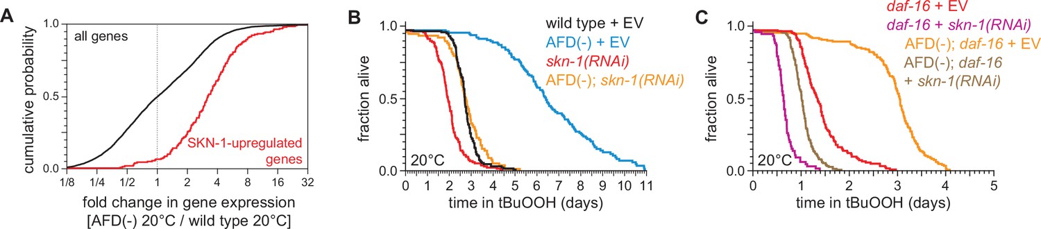
Like DAF-16, the NRF ortholog SKN-1 increases C. elegans peroxide resistance in response to reduced DAF-2 signaling (Tullet et al., 2008). The expression of genes upregulated by skn-1(+) in wild-type nematodes (Oliveira et al., 2009) and in daf-2 loss-of-function mutants (Ewald et al., 2015) was increased by AFD ablation but was not increased by higher cultivation temperature (Figure 7A and Figure 7—figure supplement 1A–C), suggesting that SKN-1 might increase peroxide resistance in AFD-ablated nematodes. Knockdown of skn-1 via RNA interference (RNAi) decreased the peroxide resistance of AFD-ablated nematodes by 58%, but caused a smaller, 27%, reduction in peroxide resistance in wild-type nematodes (Figure 7B). RNAi of skn-1 also decreased the peroxide resistance of AFD-ablated daf-16 mutants (Figure 7C). In addition, RNAi of skn-1 caused a larger reduction in peroxide resistance in daf-16 mutants when the AFD neurons were ablated than when those neurons were present (Figure 7C), suggesting that DAF-16 and SKN-1 had non-overlapping roles in promoting peroxide resistance in AFD-ablated nematodes.

Figure 7 with 1 supplement see all
SKN-1/NRF and DAF-16/FOXO collaborate to increase the nematodes’ peroxide resistance in response to signals from the AFD sensory neurons.

(A) Genes upregulated by skn-1(+) in wild-type nematodes (Oliveira et al., 2009) had higher expression in AFD-ablated nematodes grown at 20°C than in wild-type (unablated) nematodes grown at 20°C. (B) skn-1(RNAi) suppressed most of the increased peroxide resistance of AFD-ablated nematodes grown at 20°C. Control RNAi consisted of feeding the nematodes the same bacteria but with the empty vector (EV) plasmid pL4440 instead of a plasmid targeting skn-1. (C) skn-1(RNAi) lowered peroxide resistance to a greater extent in AFD-ablated daf-16(mu86) mutants at 20°C than in (unablated) daf-16(mu86) mutants at 20°C. Statistical analysis for panel (A) is in Supplementary file 3 and statistical analyses for panels (B, C) are in Supplementary file 8.

We propose that when nematodes are cultured at 20°C, the AFD neurons promote signaling by the DAF-2 insulin/IGF1 receptor in target tissues, which subsequently lowers the nematode’s peroxide resistance by repressing transcriptional activation by SKN-1/NRF and DAF-16/FOXO. However, this repression is not complete, because both daf-16(mu86) and skn-1(RNAi) lowered peroxide resistance at 20°C when the AFD neurons were present. It is also likely that DAF-16 and SKN-1 are not the only factors that contribute to peroxide resistance in AFD-ablated nematodes at 20°C, because AFD ablation increased peroxide resistance in daf-16(mu86); skn-1(RNAi) nematodes, albeit to a lesser extent than in daf-16(+) or skn-1(+) backgrounds.

DAF-16/FOXO potentiates the changes in gene expression induced by the AFD sensory neurons

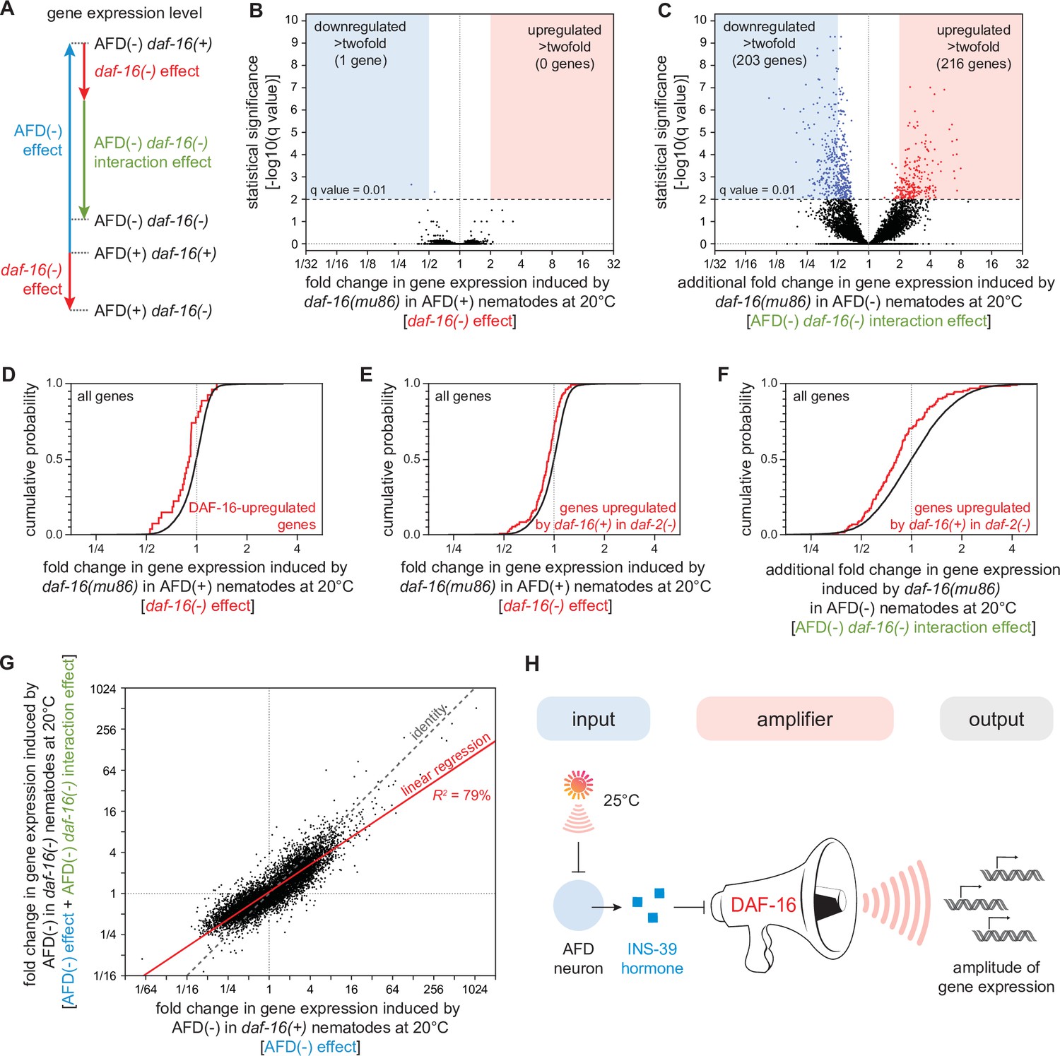
What role does the DAF-16/FOXO transcription factor play in regulating gene expression in response to signals from the AFD sensory neurons? In principle, DAF-16 could mediate all, some, or none of the changes in gene expression induced by AFD ablation. To distinguish between these possibilities, we used genome-wide epistasis analysis (Angeles-Albores et al., 2018) to compare the transcriptomes of unablated daf-16(+) [wild-type] nematodes, unablated daf-16(mu86) mutants, AFD-ablated daf-16(+) nematodes, and AFD-ablated daf-16(mu86) mutants, on day 2 of adulthood and grown at 20°C. This analysis quantified the extent to which DAF-16 affected gene expression differently in AFD-ablated and unablated nematodes (Figure 8A and Figure 8—figure supplement 1A).

Figure 8 with 2 supplements see all
DAF-16/FOXO potentiates the changes in gene expression induced by the AFD sensory neurons.

(A) We performed mRNA sequencing (mRNA-seq) on wild-type [AFD(+) daf-16(+)], daf-16(mu86) null mutants [AFD(+) daf-16(−)], AFD-ablated nematodes [AFD(−) daf-16(+)], and AFD-ablated daf-16(mu86) null mutants [AFD(−) daf-16(−)] grown at 20°C, and used an epistasis model to quantify the extent to which AFD ablation and daf-16 mutation affected the expression of each gene, relative to wild-type, in terms of the independent effects induced by AFD ablation (blue arrow) and by lack daf-16 gene function (red arrow), and the additional effect induced by the interaction between AFD ablation and lack daf-16 gene function (green arrow). Volcano plots showing the level and statistical significance of (B) the changes in gene expression induced by lack daf-16 gene function in unablated nematodes at 20°C and (C) the additional changes in gene expression induced by lack daf-16 gene function in AFD-ablated nematodes at 20°C. Genes up- and downregulated significantly (q value < 0.01) are shown in red and blue, respectively. (D, E) Effect of lack daf-16 gene function in unablated nematodes at 20°C on the expression of (C) genes directly upregulated by DAF-16 (Kumar et al., 2015) and (D) genes upregulated in a daf-16-dependent manner in daf-2(−) mutants (Murphy et al., 2003). (F) Effect of the additional changes in gene expression induced by lack daf-16 gene function in AFD-ablated nematodes at 20°C on the expression of genes upregulated in a daf-16-dependent manner in daf-2(−) mutants (Murphy et al., 2003). (G) The effect of AFD ablation on gene expression at 20°C was systematically smaller in daf-16(mu86) mutants (y-axis) than in daf-16(+) nematodes (x-axis). Linear regression fit is shown as a red line flanked by a red area marking the 95% confidence interval of the fit. (H) The DAF-16/FOXO transcription factor amplifies the changes in gene expression induced by AFD ablation. This means that DAF-16 determines the gene-expression responsiveness, but not the response to signals from the AFD sensory neurons. Statistical analyses for panels (D–F) are in Supplementary file 3.

We found that lack of daf-16 gene function in unablated nematodes at 20°C significantly lowered the expression of just two genes and significantly increased the expression of none, out of 7387 genes detected (q value <0.01) (Figure 8B). Previous transcriptomic studies in daf-2(−) mutants—unlike our study, which was conducted in daf-2(+) nematodes—have identified thousands of genes whose expression was regulated by daf-16 (Kumar et al., 2015; Lin et al., 2018; Murphy et al., 2003). We found that lack of daf-16 gene function in AFD-unablated [daf-2(+)] nematodes lowered the expression of genes directly upregulated by DAF-16 (Kumar et al., 2015; Figure 8D) and lowered the expression of genes upregulated in a daf-16-dependent manner in daf-2(−) mutants (Murphy et al., 2003; Figure 8E). However, these effects were small, averaging to just a 10% decrease in expression. These small daf-16-dependent effects on gene expression suggest that DAF-16 function was almost fully repressed by DAF-2 at 20°C. While these transcriptional effects were small, they could nevertheless contribute to the peroxide resistance of wild-type nematodes at 20°C. DAF-16 may also play a larger role in gene expression in unablated nematodes after peroxide exposure.

In contrast, when the AFD neurons were ablated, lack of daf-16 gene function induced much broader changes in gene expression, lowering the expression of 431 genes and increasing the expression of 238 genes (q value < 0.01) (Figure 8C). In addition, in AFD-ablated nematodes, lack of daf-16 lowered the expression of genes upregulated in a daf-16-dependent manner in daf-2(−) mutants (Murphy et al., 2003) to a greater degree than in unablated nematodes (Figure 8F). Taken together, these findings showed that DAF-16 played a larger role in regulating gene expression at 20°C when the AFD neurons were ablated.

Finally, we investigated how the gene-regulatory influence of the AFD neurons depended quantitatively on DAF-16. The AFD neurons and DAF-16 worked together to regulate gene expression, because the extent to which DAF-16 affected gene expression deviated systematically from the level of gene expression predicted if AFD and DAF-16 acted independently (Figure 8—figure supplement 1B). To examine how the AFD neurons and DAF-16 jointly regulated gene expression, we compared the extent to which ablation of the AFD neurons affected gene expression in daf-16(m86) mutants and in daf-16(+) nematodes. The effect of AFD ablation on gene expression was systematically smaller in daf-16(mu86) mutants than in daf-16(+) nematodes (R2 = 79%, slope = 0.67, p < 0.0001, Figure 8G). Using simulations, we showed that effect was robust despite the uncertainty in our estimates of how much AFD and DAF-16 affected the expression of each gene (see Materials and methods). In addition, we found that the extent to which AFD ablation affected the average expression of sets of genes with related functions (Higgins et al., 2022; Holdorf et al., 2020) was systematically lower in daf-16(mu86) mutants than in daf-16(+) nematodes (R2 = 86%, slope = 0.67, p < 0.0001, Figure 8—figure supplement 2). Therefore, the size of the effect of AFD ablation on gene expression was systematically smaller when the contribution of DAF-16 to gene expression was removed. We conclude that the DAF-16/FOXO transcription factor potentiates the changes in gene expression induced by ablation of the AFD sensory neurons (Figure 8H).

Discussion

Across the tree of life, organisms face the lethal threat from hydrogen peroxide attack (Avery and Morgan, 1924; Imlay, 2018). This threat is inherently temperature dependent, because the reactivity of hydrogen peroxide increases with temperature (Arrhenius, 1889; Evans and Polanyi, 1935; Eyring, 1935). In this study, we found that C. elegans nematodes use temperature information to deal with the lethal threat of hydrogen peroxide produced by the pathogenic bacterium E. faecium: when a pair of the nematodes’ neurons sensed a high cultivation temperature, they preemptively induced the nematodes’ hydrogen peroxide defenses. To our knowledge, the findings described here provide the first evidence of a multicellular organism inducing their defenses to a chemical when they sense an inherent enhancer of the reactivity of that chemical.

Temperature perception by sensory neurons regulates C. elegans hydrogen peroxide defenses

We show here that a small increase in temperature—within the range that C. elegans nematodes prefer in nature (Crombie et al., 2019)—increases the nematodes’ sensitivity to killing by environmental peroxides and by hydrogen peroxide (H2O2) produced by the pathogenic bacterium E. faecium. These effects were not due to damage to the nematodes by the higher temperature but, instead, occurred despite the nematodes inducing protective defenses in response to experiencing the higher temperature before peroxide exposure.

We found that C. elegans deals with the enhanced threat posed by environmental peroxides at high cultivation temperature by coupling the induction of their H2O2 defenses to the perception of temperature by their AFD sensory neurons. These neurons have specialized sensory endings that are the primary thermoreceptors of the nematode, enabling them to adjust their behavior and heat defenses in response to temperature (Goodman and Sengupta, 2019; Hedgecock and Russell, 1975; Prahlad et al., 2008). The AFD sensory neurons used an INS-39 insulin/IGF1 hormone—which they expressed exclusively—to relay temperature information to the intestine, the tissue that provided H2O2-protection services to the nematode. At a low cultivation temperature, AFD expressed high levels of INS-39, leading to repression of the CTL-1 and CTL-2 catalases and of other peroxide-induced genes. However, at a high cultivation temperature, AFD lowered INS-39 expression, leading to the induction of peroxide defenses by the DAF-16/FOXO transcriptional activator.

What mechanisms regulate ins-39 gene expression in the AFD neurons in response to cultivation temperature? On a short timescale of seconds to minutes, the AFD neurons respond to changes in temperature by transiently increasing intracellular [Ca2+] and changing thermoreceptor currents through a process dependent on TAX-4 cyclic GMP-gated channels (Kimura et al., 2004; Ramot et al., 2008). On a longer timescale of hours, changes in temperature can modulate gene expression within AFD through a process mediated in part by intracellular [Ca2+] via the calcium/calmodulin-dependent protein kinase CMK-1 (Ippolito et al., 2021; Yu et al., 2014). Interestingly, the baseline intracellular [Ca2+] in AFD was lower in nematodes grown continuously at 25°C than in those at 15°C, although levels at 20°C were not assessed in that work (Ippolito et al., 2021). Given that tax-4 is essential for ins-39 gene expression at both 20 and 25°C, it will be interesting to determine how cultivation temperature and TAX-4 act to regulate ins-39 gene expression in AFD on different timescales.

The repression of peroxide-protection services by the AFD neurons at the lower cultivation temperature did not rely on the neurotransmitter serotonin, unlike the induction of heat defenses by these neurons in response to 34°C heat shock (Tatum et al., 2015). AFD ablation at 20°C also induced gene sets expressed at higher levels in response to low (15°C) cultivation temperature (Gómez-Orte et al., 2018) and gene sets induced by high heat (30°C) (McCarroll et al., 2004), but those gene sets were not induced by high cultivation temperature (25°C). Therefore, the AFD sensory neurons repressed gene sets regulated by noxious heat, high cultivation temperature, and low temperature. It is possible that the AFD neurons respond to different temperature ranges by regulating the expression of specific gene sets in target tissues via different signals. In addition to expressing INS-39, these neurons express other peptide hormones—including hormones in the insulin/IGF1, FMRFamide, pigment dispersal factor, and oxytocin–vasopressin families (Barrios et al., 2012; Beets et al., 2012; Chen et al., 2016; Kim and Li, 2004; Taylor et al., 2021). It is possible that the AFD neurons also regulate the expression of specific sets of genes through temperature-independent signals; these signals could either be constitutive or regulated by other inputs sensed by AFD, such as carbon dioxide (Bretscher et al., 2011) and magnetic fields (Vidal-Gadea et al., 2015). We conclude that the AFD thermosensory neurons play a central role in the regulation of distinct systemic responses to temperature.

Target tissues control their responsiveness to sensory signals via DAF-16/FOXO

Using genome-wide epistasis analysis we showed that the DAF-16/FOXO transcription factor potentiated the changes in gene expression induced by AFD ablation at 20°C. This means that DAF-16 determined the responsiveness, but not the response, of gene expression at 20°C to signals from AFD. We reason that while C. elegans cells manage the challenge of deciding when to express specific genes by relinquishing control of that decision to signals from the AFD sensory neurons, they retain control of their responsiveness to those signals via the DAF-16/FOXO factor. Intestinal DAF-16 is also regulated by a wide variety of factors, including FLP-6 FMRFamide signals from the AFD neurons (Chen et al., 2016), insulin/IGF1 signals from sensory neurons other than AFD (Artan et al., 2016; Zhang et al., 2018), and signals from the germline (Berman and Kenyon, 2006; Hsin and Kenyon, 1999); and by signal-transduction pathways other than the insulin/IGF1 pathway, including the JNK pathway (Oh et al., 2005), AMPK pathway (Greer et al., 2007), TGFβ pathway (Liu et al., 2004; Narasimhan et al., 2011; Shaw et al., 2007), TOR pathway (Robida-Stubbs et al., 2009), and TRPA pathway (Xiao et al., 2013; Zhang et al., 2015). Therefore, we expect those factors to determine via DAF-16 the gene-expression responsiveness to signals from the AFD neurons.

Multiple types of information appear to converge in a common mechanism to regulate the induction of intestinal peroxide defenses in C. elegans. Previously, we found that the SKN-1 and DAF-16 transcription factors collaborated to mediate the induction of peroxide defenses in response to information about food levels, sensed by the ASI neurons and communicated to the intestine via a TGFβ-insulin/IGF1 hormone relay (Schiffer et al., 2020). DAF-16 and SKN-1 functions in the intestine are also regulated by signals from the germline (Berman and Kenyon, 2006; Ghazi et al., 2009; Steinbaugh et al., 2015), and TRPA-1 channels in the intestine regulate DAF-16 function in that tissue at 15°C (Xiao et al., 2013). In insects and mammals, insulin/IGF1 signaling components also regulate cellular antioxidant defenses (Brunet et al., 2004; Clancy et al., 2001; Holzenberger et al., 2003; Tatar et al., 2003). It will be interesting to explore the extent to which sensory-neuronal and other signals collaborate to regulate hydrogen peroxide defenses via insulin/IGF1 signaling in all animals.

Ideas and speculation: faithful assessment of a threat by sensing the enhancer of that threat

What does C. elegans accomplish by coupling the induction of hydrogen peroxide defenses to sensory perception of high cultivation temperature? To address this question, in this section we consider the specificity of the nematode’s strategy for dealing with the threat of hydrogen peroxide and identify the strategy’s unique features from a chemical-kinetic perspective.

How specific is the nematode’s strategy? One possibility was that temperature defenses might induce cross-protection from the stress caused by H2O2. However, high cultivation temperature (25°C) increased the expression of the intestinal catalases CTL-1 and CTL-2, which are enzymes specialized for degrading H2O2 (Amara et al., 2001; Loew, 1901; Mishra and Imlay, 2012). A second possibility was that the high cultivation temperature induced a broad set of defense responses that included those triggered by peroxides. However, that was not the case, because gene sets induced by toxic metals, organic compounds, and high-energy radiation were not induced at 25°C relative to 20°C. Therefore, the defenses induced by high cultivation temperature were specialized and included those for coping with the stress induced by hydrogen peroxide.

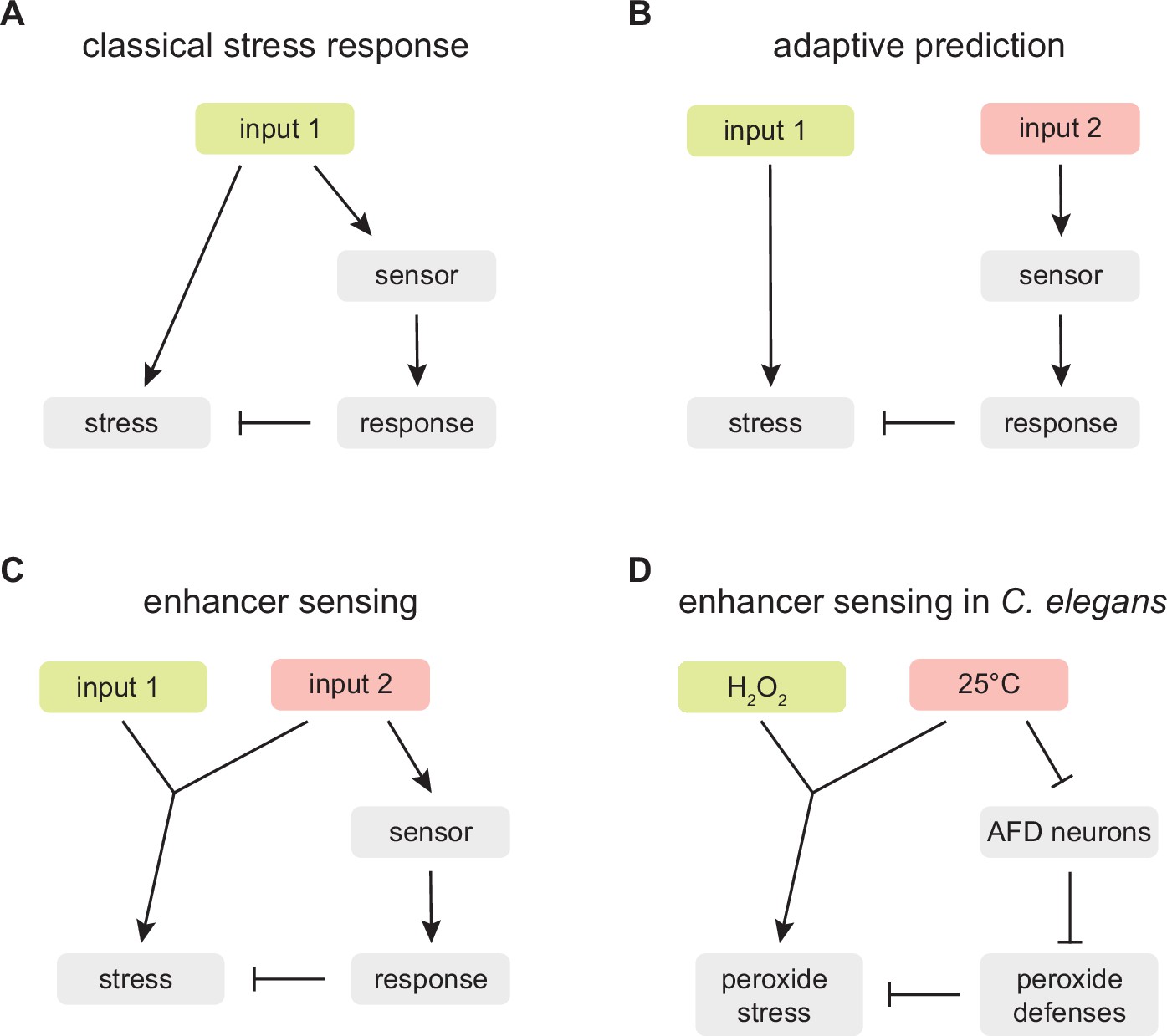
What are the strategy’s unique features? We considered the possibility that the strategy of using temperature information to preemptively induce hydrogen peroxide defenses is adaptive because when C. elegans nematodes encounter a higher cultivation temperature in their natural habitat, they are more likely to subsequently encounter H2O2 and, therefore, need H2O2 protection. Such a strategy, called adaptive prediction (Mitchell et al., 2009), is used by the bacteria E. coli and Vibrio cholerae, and by the yeasts Saccharomyces cerevisiae and Candida albicans, to sequentially induce specific defenses based on the typical order of stresses they encounter in their respective ecological settings (Mitchell et al., 2009; Rodaki et al., 2009; Schild et al., 2007; Tagkopoulos et al., 2008). Adaptive prediction would provide C. elegans with a guess of when to induce their H2O2 defenses based on how often and how quickly high temperature is followed by H2O2 exposure in C. elegans’ ecological setting (Levins, 1968; Mitchell and Pilpel, 2011).

Adaptive prediction provides a plausible explanation for why C. elegans evolved a regulatory mechanism coupling temperature perception to H2O2 defense. However, in our opinion, that explanation is insufficient, because it does not incorporate a key feature of the nematode’s strategy: the chemical constraint linking temperature and hydrogen peroxide reactivity removes guesswork from the strategy. Contrary to the expectation from adaptive prediction, C. elegans nematodes are not guessing that in their ecological setting increasing temperature leads to a higher H2O2 threat; instead, in all ecological settings the nematodes’ proteins, nucleic acids, and lipids are inherently more likely to be damaged by H2O2 with increasing temperature because those chemical reactions necessarily run faster with increasing temperature (Arrhenius, 1889; Evans and Polanyi, 1935; Eyring, 1935). This chemical constraint means that by coupling the induction of H2O2 defenses to the perception of high temperature, the nematodes are not guessing; instead, they are assessing faithfully the threat that hydrogen peroxide poses. We refer to this distinct strategy as ‘enhancer sensing’ (Figure 9).

An enhancer sensing strategy enables C.elegans to assess faithfully the threat of hydrogen peroxide using temperature information.

(A) Classical stress response: the strategy provides faithful information about the threat the organism faces because the response that enables the organism to cope with the stress induced by input 1 is coupled to the perception of input 1. (B) Adaptive prediction: the strategy provides a guess (whose predictive value matches the co-occurrence of inputs 1 and 2 in the ecological setting of the organism) but not necessarily faithful information about the threat the organism faces, because input 2 does not induce (nor affect the capacity of input 1 to induce) the stress that the organism attempts to cope with by inducing a response to input 2. (C) Enhancer sensing: the strategy provides faithful information about the threat the organism faces because the capacity of input 1 to induce a stress is modulated by input 2 and, therefore, perception of either input provides information about the threat posed by the interaction of those inputs. (D) The nematode C. elegans uses an enhancer sensing strategy that couples the de-repression of specific H2O2 defenses to the sensory perception of high temperature, an inherent enhancer of the reactivity of H2O2.

Enhancer sensing provides a new framework for understanding the adaptive value of strategies coupling the induction of defense responses to the perception of inputs that inherently modulate the need for those defenses. In a classical stress response, the strategy provides faithful information about the threat the organism faces because the response that enables the organism to cope with the stress induced by an input is coupled to the perception of that input (Figure 9A). In enhancer sensing, an input’s capacity to induce a stress is modulated by another input; the strategy provides faithful information about the threat the organism faces because perception of either input provides information about the threat posed by the interaction of those inputs (Figure 9C). In contrast, in adaptive prediction, the sensed input does not induce (nor modulate the capacity of another input to induce) the stress that the organism attempts to cope with by inducing a response to that input; as a result, the strategy does not necessarily provide faithful information about the threat the organism faces; instead the strategy provides a guess whose predictive value matches the co-occurrence in the ecological setting of the organism of the input that induces the stress and the input that is sensed (Figure 9B).

We show here that C. elegans couples H2O2 defense to the perception of high temperature. We expect this enhancer sensing strategy’s output (the level of H2O2 defense) to provide C. elegans with an evolutionarily optimal strategy across ecologically relevant inputs (cultivation temperatures) (Kussell and Leibler, 2005; Maynard, 1982; Wolf et al., 2005). This strategy is implemented at the organismic level through the division of labor between the AFD neurons, which sense and broadcast temperature information, and the intestine, which responds to that information by providing H2O2 defense (Figure 9D). Ascertaining that C. elegans uses this strategy does not depend on the temperature information broadcast by the AFD neurons exclusively regulating defense responses to temperature-dependent threats, because the regulation of defenses toward temperature-insensitive threats could affect the efficacy of defenses toward temperature-dependent threats; for example, suppressing defenses toward a temperature-insensitive threat would be beneficial if those defenses interfered with H2O2 defense or depleted energy resources contributing to H2O2 defense.

Ideas and speculation: limitations and unanswered questions

Because the studies presented here are the first to identify an enhancer sensing strategy, we do not know the extent to which this type of strategy is common across organisms. However, many previous findings may be indicative of enhancer sensing. For example, in the case of the regulation of H2O2 defenses by temperature, previous studies have shown that H2O2 defenses are induced in response to high temperature in a wide variety of organisms, including bacteria (Engelmann et al., 1995; Mossialos et al., 2006), yeasts (Deegenaars and Watson, 1997; Mitchell et al., 2009; Wieser et al., 1991), plants (Hu et al., 2021; Nishizawa et al., 2006; Panchuk et al., 2002), cnidarians (Dash and Phillips, 2012), and human HeLa cells (Pallepati and Averill-Bates, 2010).

In addition, we do not know the extent to which enhancer sensing strategies couple temperature perception to multiple defense responses within any organism. In their natural habitat, C. elegans nematodes encounter many chemicals that, like H2O2, are inherently more reactive at higher temperatures. However, it is difficult to predict whether enhancer sensing strategies coupling temperature perception to defense toward those chemicals would be likely to provide a high adaptive value because we do not know the extent to which those chemicals are common, abundant, and reactive enough to cause consequential damage within the temperature range that C. elegans experience in their ecological setting. Because growth at 25°C did not induce gene sets induced by acrylamide, formaldehyde, benzene, silver, cadmium, and arsenic, we expect that resistance to lethal concentrations of these toxic chemicals will not increase with pre-exposure to 25°C. However, it is likely that other temperature ranges sensed by the AFD neurons might regulate resistance to those chemicals, because AFD ablation at 20°C induced the gene sets induced by each of those chemicals. In the future, we plan to determine the extent to which C. elegans uses enhancer sensing strategies to couple the perception of specific temperature ranges to the induction of defenses toward these and other toxic chemicals, and whether those strategies rely on temperature perception and broadcasting by AFD and other temperature-sensing neurons. More broadly, it will be interesting to determine the extent to which enhancer sensing strategies are used throughout the tree of life to couple specific defense responses to the perception of inputs that enhance the need for those defenses.

C. elegans relies on a combination of strategies to deal with the threat of hydrogen peroxide

C. elegans decides when to induce behavioral and cellular H2O2 defenses by relying on many classes of sensory neurons (Bhatla and Horvitz, 2015; Schiffer et al., 2020; Schiffer et al., 2021). The function of these neurons can be understood in terms of their roles in different strategies enabling the nematode to deal with the lethal threat of environmental H2O2: enhancer sensing, adaptive prediction, and classical stress response. The AFD sensory neurons play a role in an enhancer sensing strategy by coupling the induction of specific H2O2 defenses to temperature perception. The ASI sensory neurons play a role in adaptive prediction by repressing H2O2 defenses in response to perception of E. coli, which protects the nematodes by depleting H2O2 from the nematode’s environment (Schiffer et al., 2020), like most bacteria in the nematodes’ ecological setting (Schiffer et al., 2021). The ASJ and I2 sensory neurons play a role in classical stress responses by triggering locomotory escape and feeding inhibition, respectively, in response to perception of H2O2 (Bhatla and Horvitz, 2015; Schiffer et al., 2021). We speculate that by relying on a combination of strategies, C. elegans nematodes can better manage the challenge of avoiding inducing costly H2O2 defenses that can cause undesirable side effects at inappropriate times.

Materials and methods

C. elegans culture, strains, and transgenes

Request a detailed protocol

Wild-type C. elegans were Bristol N2. C. elegans hermaphrodites were cultured at 20°C on NGM agar plates (Nematode Growth Medium, 17 g/l agar, 2.5 g/l Bacto Peptone, 3.0 g/l NaCl, 1 mM CaCl2, 1 mM MgSO4, 25 mM H2KPO4/HK2PO4 pH 6.0, 5 mg/l cholesterol) seeded with E. coli OP50, unless noted otherwise. Double mutant worms were generated by standard genetic methods. The Pgcy-8::ins-39(+) (pFS1) plasmid was built via gene synthesis (Twist Bioscience) by inserting the gcy-8 promoter (800 basepairs) followed by the ins-39 open reading frame and 3′ untranslated region (600 basepairs) into a pTwist High Copy vector backbone. The plasmid was injected at 30 ng/µl into ins-39(tm6467) with 50 ng/µl Punc-122::GFP as co-injection marker. ins-39(oy167[ins-39::SL2::GFP]) was made according to published protocols (Ghanta et al., 2021). Briefly, a dsDNA donor was made by amplifying SL2::GFP with 5′ SP9-modified oligos containing 35 bp overhangs homologous to the ins-39 genomic locus for insertion immediately after the stop codon, using these primers: AGCAGGTCAAAGACGACTTCGTCACACTGCTCTGAGCTGTCTCATCCTACTTTCAC (forward) and ACTGGGCAAACGGAGAGTGAACGATGGAGCATTGACTATTTGTATAGTTCATCCATGCC (reverse). The genomic locus was cut with a crRNA targeting the sequence GATGGAGCATTGATCAGAGC. For a list of all bacterial and worm strains used in this study, see Supplementary file 9 and Supplementary file 10, respectively. For a list of PCR genotyping primers and phenotypes used for strain construction, see Supplementary file 11.

Survival assays

Request a detailed protocol

Automated survival assays were conducted using a C. elegans lifespan machine scanner cluster (Stroustrup et al., 2013) as described previously (Servello and Apfeld, 2020). This platform enables the acquisition of survival curves with very high temporal resolution and large population sizes. All chemicals were obtained from Sigma. For survival assays with 1 mM hydrogen peroxide and 6 mM tert-butyl hydroperoxide, the respective compound was added to molten agar immediately before pouring onto 50 mm NGM agar plates. Plates were dried (Stroustrup et al., 2013) and seeded with 100 µl of concentrated E. coli OP50 resuspended at an OD600 of 20 (Entchev et al., 2015). For RNAi experiments, the appropriate E. coli HT115 (DE3) strain was used instead. For hydrogen peroxide assays, E. coli JI377 was used instead (Seaver and Imlay, 2001). Nematodes were cultured at the specified developmental temperature until the onset of adulthood, and then cultured at the specified adult temperature, in groups of up to 100, on plates with 10 μg/ml 5-fluoro-2′-deoxyuridine (FUDR), to avoid vulval rupture (Leiser et al., 2016) and eliminate live progeny. In a previous study (Schiffer et al., 2020), as an alternative to FUDR, we inhibited formation of the eggshell of fertilized C. elegans embryos with RNAi of egg-5 (Entchev et al., 2015), with identical results in wild-type nematodes and daf-7(−) mutants, which increase peroxide resistance. Day 2 adults were transferred to lifespan machine assay plates. A typical experiment consisted of up to 4 genotypes or conditions, with 4 assay plates of each genotype or condition, each assay plate containing a maximum of 40 nematodes, and 16 assay plates housed in the same scanner. All experiments were repeated at least once, yielding the same results. Scanner temperature was calibrated to 20 or 25°C with a thermocouple (ThermoWorks USB-REF) on the bottom of an empty assay plate. Death times were automatically detected by the lifespan machine’s image-analysis pipeline, with manual curation of each death time through visual inspection of all collected image data (Stroustrup et al., 2013), without knowledge of genotype or experimental condition.

E. faecium survival assays

Request a detailed protocol

Nematodes were continuously fed E. coli JI377 for at least three generations and grown under the specified developmental temperature until the onset of adulthood, then cultivated at the specified adult temperature on plates with 10 μg/ml FUDR. E. faecium E007 was cultured in 500 ml of Brain Heart Infusion (BHI) medium overnight at 37°C without aeration and then aerated for 4 hr at 37°C prior to collecting the supernatant. The supernatant was stored at −20°C and incubated at the specified temperature before use. Nematodes were washed with M9 buffer with 0.01% Tween and transferred into 24-well plates containing either BHI or the appropriate amount of E. faecium E007 supernatant, E. coli JI377 at an OD600 of 2, in a final volume of 2 ml. Plates were sealed with parafilm, incubated at the specified temperature, and survival was scored after 16 hr by mixing the wells via pipette and scoring for movement using a dissection stereo microscope equipped with white-light transillumination.

Transcriptomic analysis

Request a detailed protocol

mRNA for sequencing was extracted from day 2 adult animals. Worms were cultured on NGM agar plates seeded with E. coli OP50 and synchronized at the late L4 stage by transfer onto new NGM agar plates seeded with E. coli OP50 and supplemented with 10 μg/ml FUDR. Worms were cultured at 20°C except for the growth temperature assay for which worms were cultured at 25 or 20°C for four generations before sampling at the respective temperatures. We adapted a nematode lysis protocol (Ly et al., 2015) for bulk lysis to pool 30 individuals per sample in 120 µl of lysis buffer. cDNA preparation from mRNA was performed by SmartSeq2 as described (Picelli et al., 2014). cDNA was purified using an in-house paramagnetic bead-based DNA purification system mimicking Agencourt AMPure XP magnetic beads. Dual-barcoded Nextera sequencing libraries were prepared according to the manufacturer’s protocol and purified twice with magnetic beads. Libraries were sequenced on an Illumina NextSeq 500 with a read length of 38 bases and approx. 2.0 × 10⁶ paired-end reads per sample. RNA-seq reads were aligned to the C. elegans Wormbase reference genome (release WS265) using STAR version 2.6.0c (Dobin et al., 2013) and quantified using featureCounts version 2.0.0 (Liao et al., 2014), both using default settings. To quantify the expression within intervals in the genomic region encoding the three C. elegans catalase genes, we created a GTF that matches genomic positions defined previously (Petriv and Rachubinski, 2004). The reads count matrix was normalized using scran (Lun et al., 2016). Differential analysis was performed using a negative binomial generalized linear model as implemented by DESeq2 (Love et al., 2014). A batch replicate term was added to the regression equation to control for confounding. Batch-corrected counts were obtained by matching the quantiles of distributions of counts to the batch-free distributions as in the Combat-seq method (Zhang et al., 2020). Principal component analysis was performed on the batch-corrected and normalized log counts with a pseudo-count of one. To access the significance of the slope between the sum of the ‘AFD(−) effect’ and ‘AFD(−) daf-16() interaction effect’ coefficients and the ‘AFD(−) effect’ coefficient, we simulated 1000 sets of coefficients using a normal distribution with mean equal to the maximum-likelihood estimate of the coefficients and with standard deviation equal to the standard error of the estimates. We then fit a linear regression to each of the simulated coefficients and computed their coefficient of determination (R2). These simulations showed that, after accounting for the level of uncertainty on our estimates of the values of the coefficients for ‘AFD(−) effect’ and ‘AFD(−) daf-16() interaction effect’, the average value of the regression’s slope was 0.6826 (99% confidence interval [0.6825, 0.6827]) and the average R2 was 0.699 (99% confidence interval [0.6989, 0.6991]). GO enrichments, tissue enrichment analysis, and phenotypic enrichment analysis were determined by using the WormBase Enrichment Suite (Angeles-Albores et al., 2016). We clustered and plotted GO terms with q value <10−6 using REVIGO (Supek et al., 2011). Curated gene-expression datasets were obtained from WormExp (Yang et al., 2016). A curated hierarchical classification of genes into sets based on physiological function, molecular function, and cellular location was obtained from WormCat (Higgins et al., 2022; Holdorf et al., 2020).

Microscopy

Request a detailed protocol

Nematodes carrying the ins-39(oy167[ins-39::SL2::GFP]) allele were immobilized with 20 mM tetramisole, mounted on 10% agarose pads on slides, and imaged on a Zeiss Axio Imager M2 epifluorescent microscope with a ×63 objective. Exposure time was set to 300 ms and images were acquired with 2 × 2 binning. Detectable GFP expression was determined to be entirely restricted to AFD, in day 1 adults, by coexpression of the AFD-specific marker ttx-1p::mCherry (Satterlee et al., 2001). Images for GFP quantification were acquired with no red marker in the background. Images were processed in ImageJ, and expression was quantified from a maximum projected z-stack as corrected total cell fluorescence (CTCF) by the equation: CTCF = integrated density − (area of selected cell ROI × mean fluorescence of a nearby background ROI).

RNA interference

Request a detailed protocol

E. coli HT115 (DE3) bacteria with plasmids expressing dsRNA targeting specific genes were obtained from the Ahringer and Vidal libraries (Kamath et al., 2001; Rual et al., 2004). Empty vector plasmid pL4440 was used as control. Bacterial cultures were grown in LB broth with 100 μg/ml ampicillin at 37°C and seeded onto NGM agar plates containing 50 μg/ml carbenicillin and 2 mM IPTG. Nematodes were cultivated on E. coli OP50 until day 2 of adulthood, then transferred to RNAi plates. Their progeny was subsequently used for each assay.

Kinetic modeling

Request a detailed protocol

The Eyring–Polanyi equation, derived from transition state theory, describes how chemical reactions depend on temperature (Evans and Polanyi, 1935; Eyring, 1935):

(1) k=κkBTheSRe-HRT

where k is the rate coefficient of the reaction, T is the temperature, ∆H is the enthalpy of activation of the reaction, ∆S is the entropy of activation of the reaction, R is the ideal gas constant, kB is the Boltzmann constant, h is the Plank constant, and κ is the transmission coefficient. The relative rate of a reaction at two different temperatures is the ratio of the reaction’s rate coefficients at those temperatures:

(2) k1k2=T1T2e-HR1T1-1T2

We obtained the ∆H values of specific reactions of H2O2 from published data: 46.6 kJ/mol for the Fenton reaction with DNA-bound Fe(II) at neutral pH in the presence of ATP (Park et al., 2005), 20 kJ/mol for the thiol oxidation of the highly reactive catalytic cysteine of alkyl hydroperoxide reductase E from Mycobacterium tuberculosis (Zeida et al., 2014), between 20 and 65 kJ/mol for the thiol oxidation of the reactive cysteine residue in (the non-hydroperoxidase) glyceraldehyde-3-phosphate dehydrogenase (GAPDH) (Deponte, 2017), above 100 kJ/mol for the thiol oxidation of regular cysteines in proteins (Deponte, 2017), and 68.5 kJ/mol for the thiol oxidation of free cysteine in aqueous solution (Luo et al., 2005).

Statistical analysis

Request a detailed protocol

Statistical analyses were performed in JMP Pro version 15 (SAS). Survival curves were calculated using the Kaplan–Meier method. We used the log-rank test to determine if the survival functions of two or more groups were equal. We used analysis of variance (ANOVA) to determine whether the fold-change in gene expression of specific gene sets and of all genes were equal. We used ANOVA for GFP expression comparisons and, in cases where more than two groups were compared, used the Tukey HSD post hoc test to determine which pairs of groups in the sample differed. We used the Cell chi-square test to determine if a cell in a table differed from its expected value in the overall table. We used ordinal linear regression to determine whether the proportions of dead animals after treatment with E. faecium supernatant were equal across groups and to quantify interactions between groups using the following linear model: data = intercept + group 1 + group 2 + group 1 × group 2 + ε. The second to last term in this model quantifies the existence, magnitude, and type (synergistic or antagonistic) of interaction between groups. We used the Bonferroni correction to adjust p values when performing multiple comparisons.

Materials availability

Request a detailed protocol

Further information and requests for resources and reagents should be directed to and will be fulfilled by the Lead Contact, Javier Apfeld (j.apfeld@northeastern.edu).

Data availability

Raw mRNA-seq read files are available under Bioproject PRJNA822361 (https://www.ncbi.nlm.nih.gov/bioproject/PRJNA822361). All data generated or analyzed during this study are included in the manuscript and supporting files.

The following data sets were generated
    1. Apfeld J
    (2022) NCBI BioProject
    ID PRJNA822361. Neuronal temperature perception induces specific defenses that enable C. elegans to cope with the enhanced reactivity of hydrogen peroxide at high temperature.

References

    1. Wieser R
    2. Adam G
    3. Wagner A
    4. Schüller C
    5. Marchler G
    6. Ruis H
    7. Krawiec Z
    8. Bilinski T
    (1991)
    Heat shock factor-independent heat control of transcription of the CTT1 gene encoding the cytosolic catalase T of Saccharomyces cerevisiae
    The Journal of Biological Chemistry 266:12406–12411.

Decision letter

  1. Douglas Portman
    Reviewing Editor; University of Rochester, United States
  2. Claude Desplan
    Senior Editor; New York University, United States
  3. Douglas Portman
    Reviewer; University of Rochester, United States

In the interests of transparency, eLife publishes the most substantive revision requests and the accompanying author responses.

Decision letter after peer review:

Thank you for submitting your article "Neuronal temperature perception induces specific defenses that enable C. elegans to cope with the enhanced reactivity of hydrogen peroxide at high temperature" for consideration by eLife. Your article has been reviewed by 3 peer reviewers, including Douglas Portman as Reviewing Editor and Reviewer #1, and the evaluation has been overseen by Claude Desplan as the Senior Editor.

The reviewers have discussed their reviews with one another, and the Reviewing Editor has drafted this to help you prepare a revised submission.

Essential revisions:

1) Your model proposes but does not directly test the idea that the requirement for AFD in 25C-induced peroxide resistance reflects a role for thermosensation by AFD in this process. In your revision, please test this directly (for example, using gcy-18 gcy-8 gcy-23 triple mutants in which AFD thermosensation is abolished) or, at a minimum, determine whether AFD activity is required (for example, by silencing AFD or by rescuing tax-4 in AFD).

2) Your model does not directly address whether AFD is the relevant site of ins-39 function. In your revision, please test this directly by AFD-specific rescue and/or disruption of ins-39.

3) In several places in the paper, the reviewers feel that your paper oversimplifies the role of daf-16 in temperature-induced peroxide resistance. More detail about these concerns can be found in the individual reviews below. Please edit the manuscript to more clearly address AFD-independent roles of daf-16 as well as daf-16/skn-1-independent roles of AFD.

4) The reviewers feel that the "enhancer sensing" idea, while intriguing, is not supported strongly enough by the paper's results. Please carry out additional experiments (as suggested by reviewers 1 and 3) and/or tone down the writing to make it clear that this idea is speculative. Please also consider Reviewer 3's concern about potential confusion caused by the use of the term "enhancer sensing".

Each reviewer has additional comments and suggestions below. While we ask that you consider all of these, it is not necessary for your revision to include new experiments to address these points.

Reviewer #1 (Recommendations for the authors):

Here, Servello et al., explore the role of temperature and the temperature-sensing neuron AFD in promoting protection against peroxide damage. Unlike many other environmental threats, peroxide toxicity is expected to be temperature-dependent, since its chemical reactivity should be enhanced by higher temperatures. The authors convincingly and rigorously show that transient exposure to 25C, a condition of mild heat stress in C. elegans, activates animals' defenses against peroxides but potentially not other agents. Interestingly, this response requires the temperature-sensing AFD neurons, though whether temperature-dependent AFD activity is itself involved in this regulation is not explored. Further, the authors find that temperature regulates AFD's expression of the insulin ins-39 and provide evidence supporting the idea that repression of ins-39 at 25C contributes to enhanced peroxide defense. The authors use transcriptomic approaches to explore gene expression changes in animals in which AFD neurons are ablated, providing evidence that the FoxO-family transcription factor DAF-16 potentiates AFD signaling. However, because AFD ablation triggers effects broader than transient 25C exposure, the significance of these findings for temperature-dependent peroxide defense is somewhat unclear. Additionally, the possibility that DAF-16 (as well as another protective factor, SKN-1) function in parallel to temperature stress is consistent with many of the results shown but is not as thoroughly considered. Together, these studies identify a fascinating example of pre-emptive threat response triggered by the detection of a potentiator of that threat, a phenomenon they term "enhancer sensing." While some predictions of the specificity of this phenomenon remain untested, the paper provides intriguing insight into the potential mechanisms by which it may occur.

The dependence of the enhancer-sensing phenomenon on AFD leads the authors to conclude that the 25C stimulus is sensed by AFD itself, but this needs to be directly tested. To do this, they could ask whether tax-4 function is required in AFD, or use mutants in which AFD's thermosensory function is compromised.

The enhancer-sensing model is fascinating, but as it stands it is somewhat oversold. The authors could tone down the writing, indicating that this model is suggested rather than shown. Alternatively, they could more carefully test some of its predictions – for example by exploring the response to other threats (e.g. some of the toxicants described in Figure S5) at 20C and 25C in WT and AFD-ablated animals.

The role of ins-39 remains somewhat speculative. Figure 4F shows that ins-39 mutants have a reduced induction of peroxide defense, but it seems that this could be the result of a ceiling effect. The authors' model predicts that overexpression of ins-39, particularly at 25C, should sensitize animals to peroxide damage, a prediction that should be tested directly. Further, the authors seem to assume that AFD is the relevant site of ins-39 function, but this needs to be better supported.

Most of the daf-16 and skn-1 experiments are carried out in AFD-ablated animals, making the relevance of these findings for the 25C-dependent induction of peroxide defense somewhat unclear. As the authors show, AFD ablation causes much more extensive changes than transient 25C exposure, clearly seen in slope of the line in 3C. Further, unlike 25C exposure, AFD ablation is a chronic and non-physiological state. It would be useful for the authors to be cautious in their interpretation of these findings and to be clearer about how strongly they can connect them to the "enhancer sensing" phenomenon. Along these lines, the potentiation idea could be toned down a bit. Much of the data is consistent with parallel function for daf-16 (and skn-1) – for example, Figure 5C indicates additive effects of daf-16 and 25C exposure; 6C shows that AFD ablation still has a clear effect on peroxide sensitivity in the absence of both daf-16 and skn-1; and Figure S8a shows that much of the transcriptional response to AFD ablation (along PC1) is intact in daf-16 animals.

Based on theory or data, it would be useful for the authors to be more specific about the extent to which a 5-degree rise in temperature would be expected to enhance peroxide damage. The idea itself is solid, but whether the size of the effect is large enough to be biologically meaningful isn't addressed.

FUdR is used in all of the peroxide-sensitivity assays. Is there any reason to be concerned about this? It would be useful for the authors to comment on the reasons why they don't (or do?) expect disruption of germline proliferation to influence responses to peroxide/temperature/AFD ablation.

In a number of places, I think the writing could be toned down a bit. Some examples:

Line 124 – authors use the word multiple, but unless I've missed something, here this means two. "Multiple" is not inaccurate but is a bit misleading.

Line 154-55 – these are empirical observations, so the idea that the worm is changing its physiology "to prepare for" something is speculative.

Lines 181-2 – to me, these results suggest that neuronal sensory transduction by tax-4 channels is important.

Line 294 – because AFD ablation isn't a physiological state, I'd suggest avoiding the suggestion that defenses are "pre-induced"

One stylistic comment that the authors should feel free to ignore and do not need to address in their rebuttal: I find the repeated use of "the nematodes" in the text to be a little strange. Referring instead to "C. elegans" or "animals" would be a more standard approach.

Reviewer #2 (Recommendations for the authors):

In this study, Servello and the colleagues characterize how a temperature sensing neuron AFD regulates increased resistance to hydrogen peroxide in worms cultivated at a higher temperature. They show that loss of AFD and the insulin-like peptide INS-39 produced by AFD increase H2O2 resistance similarly as high temperature growth. To understand the molecular basis, they use mRNA-seq and analysis of gene expression at the whole-genome scale and transgenic lines to show that AFD ablation and high cultivation temperature generate overlapping changes in gene expression via the function of the FOXO transcription factor DAF-16 in the intestine.

This study is built on their previous work that established C. elegans as a model to study mechanisms for sensing and resistance of H2O2, an important environmental chemical threat for living organisms. Here, the authors uncover the neuronal and molecular basis for H2O2 resistance induced by high cultivation temperature. The authors use multiple approaches, including genetics, transgenics, whole-genome gene expression analysis, to characterize "enhancer sensing" that they discovered in this study. The experiments are well designed with appropriate controls. The data analysis is comprehensive and revealing. The findings are novel and explain a common and interesting phenomenon. The new understanding generated in this study will appeal to the readers in the fields of sensory biology, signaling transduction and physiology. The implications or conclusions of a few results presented here could be further discussed or clarified in the context of several previous studies.

1. My main question is about the link between AFD's response to higher temperature, its activity, and ins-39 expression. Previous studies show that increasing temperature from 20 to 25 degree activates AFD measured by intracellular calcium imaging. These results together with the findings in this paper would suggest that increased AFD activity reduces ins-39 expression. It will be helpful for the authors to discuss about these implications more clearly. In this paper, the authors seem to suggest that higher temperature at 25 inhibits AFD to reduce ins-39 expression. This may lead to the prediction that in the tax-4 mutant, in which AFD is not active in response to temperature, ins-39 expression is higher than wild type. This is different from the results in Figure 4E. It is possible that the effect of growing at 25 degree on AFD is different from acute sensing of the higher temperature. It will be much helpful for authors to discuss and clarify these points clearly.

2. Figure 1. Is H2O2 similarly stable at 20 degree and 25 degree?

3. Fig2H shows that AFD ablation has a stronger effect in increasing resistance than growing at 25c -- the additional effect caused by AFD ablation could use some discussion.

4. The rationale for the authors to focus on ins-39 needs to be better clarified, since multiple INSs are found in AFD.

5. Figure 4F. When growing at 25c, no further increase in resistance is seen in the ins-39 mutant compared to wild type, indicating a full effect; but at 20c, the ins-39 mutant does not fully mimic the resistance in worms grown at 25c. How would the authors explain this partial effect?

6. Figure 4G. CenGEN also identified the expression of ins-39 in several other neurons including ASK and ASJ. Therefore, the AFD-specific function of ins-39 in regulating H2O2 resistance should be further clarified.

7. Figure 3G. The authors suggest that increased ctl-1 and ctl-2 expression in AFD(-) worms confers increased resistance, because overexpression of these genes increases H2O2 resistance as shown in previous studies. This analysis of ctl genes provide evidence for the mechanistic basis for increased H2O2 resistance in AFD(-) worms. Is the increased expression of ctl-1 and ctl-2 in AFD(-) worms comparable to those generated by overexpression (ie does it confer higher resistance)? Some level of support or clarification will be very helpful.

8. Figure 7. It is much easier to read if most of the analysis in FigS8 is included in Fig7.

Reviewer #3 (Recommendations for the authors):

This paper offers novel mechanistic insights into how pre-exposure to warm temperature increases the resistance of C. elegans to peroxides, which are more toxic at warmer temperature. The temperature range tested in this study lies within the animal's living conditions and is much lower than that of heat shock. Therefore, this study expands our understanding of how past thermosensory experience shapes physiological fitness under chemical stress. The paper is technically sound with most experiments or analyses carried out rigorously, and therefore the conclusions are solid. However, it challenges our current understanding of the role of the C. elegans thermosensory system in coping with stress. The traditional view is that the AFD thermosensory neuron is activated upon sensing temperature rise, and that temperature sensation through AFD positively regulates systemic heat shock response and promotes longevity in C. elegans. Thus, it is quite unexpected that AFD ablation activates DAF-16 and improves peroxide resistance. It also appears counterintuitive that genes upregulated at 25 degrees overlap extensively with those upregulated by AFD ablation at 20 degrees. I feel that it is premature to coin the term "enhancer sensing" for such a phenomenon, as their work does not rule out the possibility that AFD ablation increases resistance to other stresses that are independent of temperature regarding their toxicity or magnitude of hazard. Additional work is necessary to clarify these issues.

1. Whether the role of AFD in inhibiting peroxide resistance is related to AFD activity needs further clarification. AFD activity depends on the animal's thermosensory experience. As animals in this study are maintained at 20 degrees unless indicated specifically, the AFD displays activities starting around 17 degrees and peaks around 20 degrees. Under such condition, the AFD displays little or no activity to thermal stimuli around 15 degrees. It will be important to test whether cultivation of animals at 20 degrees improves peroxide resistance at 15 degrees, compared to 15 degrees-cultivation/15 degrees peroxide testing. The authors should also test whether AFD ablation further improves survival under peroxides at 15 degrees for animals grown at 20 degrees, whose AFD should show little or no activities at 15 degrees.

2. The importance of the thermosensory function of AFD should be verified. In the current study, the tax-4 mutation was used to infer AFD activity, but tax-4 is expressed in sensory neurons other than AFD. In addition to AFD, AWC can sense temperature and it also expresses tax-4. Therefore, influence on AFD from other tax-4-expressing neurons cannot be excluded. On the other hand, ablation of AFD removes all AFD functions, including those that are constitutive and temperature-independent. Therefore, the authors should test the gcy-18 gcy-8 gcy-23 triple mutant, in which the AFD neurons are fully differentiated but completely insensitive to thermal stimuli. These three thermosensor genes are exclusively expressed in AFD. Compared to the tax-4 mutant that is broadly defective in multiple sensory modalities, this triple gcy mutant shows defects specifically in thermosensation. They should see whether results obtained from the AFD ablated animals could be reproduced by experiments using the gcy-18 gcy-8 gcy-23 triple mutant. The authors are also recommended to investigate ins-39 expression in AFD and profile gene expression patterns in the gcy-18 gcy-8 gcy-23 triple mutant.

3. The literature suggests that AFD promotes longevity likely in part through daf-16 (Chen at al., 2016) or independent of daf-16 (Lee and Kenyon, 2009). Whatever it is, various studies show that activation of AFD and daf-16 promote a normal lifespan at higher temperature, and AFD ablation shortens lifespan at either 20 or 25 degrees. Therefore, the finding that DAF-16-upregulated genes overlap extensively with those upregulated by AFD ablation is quite unexpected (Figure 5B). The authors should perform further gene ontology (GO) analysis to identify subsets of genes co-regulated by DAF-16 and AFD ablation, whether these genes are reported to be involved in longevity regulation, immunity, stress response, etc.

4. I feel that "enhancer sensing" is an overstatement, or at least a premature term that is not sufficiently supported without further investigations. The authors should explore whether AFD ablation or pre-exposure to warm temperature specifically enhances resistance to a stressor the toxicity of which is increased at higher temperature, but does not affect the resistance to other temperature-insensitive threats.

5. The authors need to provide data of all survival assays as supplemental tables, and indicate the sets of data that are selected to be presented in the figures. This is standard practice for C. elegans lifespan or survival assays and is important for the readers to understand the reproducibility of the experiments. This applies to Figures 1A, 1C, 1D, 2B-E, 2H, 4F, 5C-F, 6B, 6C, S1A, S1B, S2, S6B and S7C.

6. Page 17, Line 437-438: Loss of daf-16 in AFD-intact animals results in changes in the expression of merely two genes, which is quite surprising given the critical role of daf-16 in lifespan and stress resistance. Although the authors explained this in Discussion saying that fold changes in most genes were mild in the daf-16(mu86) mutant, it might be helpful to point out that in previous studies, many daf-16-dependent genes were characterized by comparing gene expressions in the daf-2 and daf-16; daf-2 double mutants. Given the critical importance of daf-16 in fitness, stress resistance and longevity, this apparent discrepancy with the existing literature must be explained in sufficient detail.

7. DAF-16 and SKN-1 nuclear localization should be investigated at warm temperature, in the absence of AFD and in the ins-39 mutant.

8. Figure 3G: The fold change in the expression levels of ctl-1, ctl-2 and ctl-3 at 25 degrees (v.s. 20 degrees) and in AFD-ablated animals should be confirmed by RT-qPCR. The data from RNA-seq in their current form do not allow for statistical analysis to determine whether the expression of these ctl genes are significantly upregulated by high cultivation temperature or AFD ablation.

9. It would be interesting to characterize gene expression changes in the ins-39 mutant and make comparison to those induced by temperature shift and AFD ablation. However, I understand that this is a substantial amount of work and it is OK if the authors decide not to do it at this moment.

10. Please provide statistics in all the Figure panels instead of using designations such as "a, b, c".

11. For esthetical reasons, the authors should remove the black portions in Figure 4B that were used to rectify the borders of the rotated fluorescent images. However, I think there is still room for careful cropping of the fluorescent images to make them rectangular without having to add another layer of black background.

12. Page 13, Line 327: please describe the molecular lesion of ins-39(tm6467).

13. Please indicate sample sizes for experiments in Figure 4C and 4D.

14. Remove the "A" label for Figure S2.

[Editors’ note: further revisions were suggested prior to acceptance, as described below.]

Thank you for resubmitting your work entitled "Neuronal temperature perception induces specific defenses that enable C. elegans to cope with the enhanced reactivity of hydrogen peroxide at high temperature" for further consideration by eLife. Your revised article has been evaluated by Claude Desplan (Senior Editor) and a Reviewing Editor.

All of the reviewers feel that your manuscript has been substantially improved. However, in their discussion, the reviewers continued to express concerns about the validity of the "enhancer sensing" model. Before publication, we ask that you revise the Abstract and Discussion to present a more balanced view. Any other changes that you may wish to make in response to the comments below are optional.

Reviewer #1 (Recommendations for the authors):

In their revised manuscript, Servello et al., have nicely addressed most of the concerns I raised earlier. I find the manuscript to be significantly improved. Before the paper is ready for publication, I think it would be worthwhile for the authors to consider addressing the following issues by editing the text.

– The demonstration that the gcy triple mutant has a peroxide-resistant phenotype is nice, but it would be useful for the authors to speculate about why the phenotype of this mutant is so much weaker than ADF ablation. The phenotypes of the single gcy mutants are also a little unexpected; this should be noted somewhere.

– I continue to feel that the "enhancer sensing" model is pushed too hard. In particular, I find the argument in the Discussion, lines 657 to 688, to be unconvincing. Adaptive sensing provides a mechanism by which species can "learn" to associate one piece of information with another, allowing detection of a particular environmental signal to become predictively coupled to an apparently distinct response. Here, the authors have shown (quite nicely) that C. elegans has "learned" that an increase in temperature predicts an increased susceptibility to peroxide damage. To me, this fits quite well with the adaptive sensing paradigm. I'm ok with the idea that what they are seeing could be considered a variant form of adaptive sensing, but it seems to me to be excessive to decide that a new term is needed for what amounts to essentially the same principle. In a way, even that is overkill: because the hazard is the thermodynamic effect of temperature itself, one could argue that increasing peroxide defense is an aspect of temperature defense, in the same way that increasing expression of protein chaperones is. As the authors note, it's well demonstrated that heat detection by the C. elegans nervous system can trigger the systemic activation of the heat shock response. The activation of peroxide defenses seems to me to be, in principle, an equivalent phenomenon.

– The authors write in their rebuttal that it is incorrect to predict that the response to non-H2O2 stresses should not be enhanced by 25C pre-exposure. In principle, I agree – one can't necessarily predict that augmenting defenses against these stressors wouldn't also be adaptive at 25C – but isn't the point of lines 340-342 to indicate that pre-exposure to 25C does indeed have some selectivity in terms of the stress responses it enhances? If so, then there seems to be a strong prediction that animals' responses to acrylamide, formaldehyde, etc, would not be changed by 25C pre-exposure.

Reviewer #3 (Recommendations for the authors):

The authors addressed most of the points that I raised in the initial review of the study. Although I am still not completely convinced by the idea of enhancer sensing and some of the interpretations of the role of AFD on gene expression and physiology, in particular its interaction with daf-16, I think those issues are better addressed in a separate study. Regarding "enhancer censing," for such a new term to be coined, multiple examples are necessary. Therefore, I suggest that the authors tone down this claim in the Discussion/Abstract, and also discuss the potential limitations of their study in justifying such a claim. In short, a more balanced view is needed.

https://doi.org/10.7554/eLife.78941.sa1

Author response

Essential revisions:

1) Your model proposes but does not directly test the idea that the requirement for AFD in 25C-induced peroxide resistance reflects a role for thermosensation by AFD in this process. In your revision, please test this directly (for example, using gcy-18 gcy-8 gcy-23 triple mutants in which AFD thermosensation is abolished) or, at a minimum, determine whether AFD activity is required (for example, by silencing AFD or by rescuing tax-4 in AFD).

As requested, we determined whether previously identified mechanisms for temperature perception by the AFD neurons were required for the temperature-dependent regulation of peroxide resistance using gcy-18 gcy-8 gcy-23 triple mutants and the respective single mutants. The findings from the new experiments lead us to conclude that temperature perception by AFD via GCY-8, GCY-18, and GCY-23 enables C. elegans to lower their peroxide resistance at the lower cultivation temperature.

2) Your model does not directly address whether AFD is the relevant site of ins-39 function. In your revision, please test this directly by AFD-specific rescue and/or disruption of ins-39.

As requested, we determined whether ins-39 gene expression in the AFD neurons was sufficient to lower peroxide resistance by restoring ins-39(+) gene expression only in the AFD neurons using the AFD-specific gcy-8 promoter. The findings from this experiment lead us to conclude that expression of ins-39 in the AFD neurons was sufficient to regulate C. elegans peroxide resistance.

3) In several places in the paper, the reviewers feel that your paper oversimplifies the role of daf-16 in temperature-induced peroxide resistance. More detail about these concerns can be found in the individual reviews below. Please edit the manuscript to more clearly address AFD-independent roles of daf-16 as well as daf-16/skn-1-independent roles of AFD.

We have edited the manuscript to more clearly address the roles of DAF-16 in the response of peroxide resistance to cultivation temperature, and the roles of DAF-16 and SKN-1 when the AFD neurons are present and when they are ablated, as detailed in our responses to reviewer #1’s point 4 and to reviewer #3’s point 3 and additional issue 2.

4) The reviewers feel that the "enhancer sensing" idea, while intriguing, is not supported strongly enough by the paper's results. Please carry out additional experiments (as suggested by reviewers 1 and 3) and/or tone down the writing to make it clear that this idea is speculative. Please also consider Reviewer 3's concern about potential confusion caused by the use of the term "enhancer sensing".

To address these concerns, we edited the manuscript and expanded the manuscript’s discussion, as detailed in our responses to reviewer #1’s point 2 and to reviewer #3’s point 4. The revised manuscript now makes a clear distinction between (a) enhancer sensing as a strategy for the regulation of defense responses (a conceptual advance we introduce in this paper) and (b) the specific instance of enhancer sensing we found is used by C. elegans to couple the induction of H2O2 defenses to the perception of temperature. Additionally, we now (c) discuss the evolutionary contexts that may favor or constrain the evolution of enhancer sensing strategies coupling temperature perception to the induction of defenses towards reactive chemicals other than hydrogen peroxide.

Reviewer #1 (Recommendations for the authors):

Here, Servello et al., explore the role of temperature and the temperature-sensing neuron AFD in promoting protection against peroxide damage. Unlike many other environmental threats, peroxide toxicity is expected to be temperature-dependent, since its chemical reactivity should be enhanced by higher temperatures. The authors convincingly and rigorously show that transient exposure to 25C, a condition of mild heat stress in C. elegans, activates animals' defenses against peroxides but potentially not other agents. Interestingly, this response requires the temperature-sensing AFD neurons, though whether temperature-dependent AFD activity is itself involved in this regulation is not explored. Further, the authors find that temperature regulates AFD's expression of the insulin ins-39 and provide evidence supporting the idea that repression of ins-39 at 25C contributes to enhanced peroxide defense. The authors use transcriptomic approaches to explore gene expression changes in animals in which AFD neurons are ablated, providing evidence that the FoxO-family transcription factor DAF-16 potentiates AFD signaling. However, because AFD ablation triggers effects broader than transient 25C exposure, the significance of these findings for temperature-dependent peroxide defense is somewhat unclear. Additionally, the possibility that DAF-16 (as well as another protective factor, SKN-1) function in parallel to temperature stress is consistent with many of the results shown but is not as thoroughly considered. Together, these studies identify a fascinating example of pre-emptive threat response triggered by the detection of a potentiator of that threat, a phenomenon they term "enhancer sensing." While some predictions of the specificity of this phenomenon remain untested, the paper provides intriguing insight into the potential mechanisms by which it may occur.

The dependence of the enhancer-sensing phenomenon on AFD leads the authors to conclude that the 25C stimulus is sensed by AFD itself, but this needs to be directly tested. To do this, they could ask whether tax-4 function is required in AFD, or use mutants in which AFD's thermosensory function is compromised.

We thank the reviewer for suggesting these experiments. As requested, we determined whether previously identified mechanisms for temperature perception by the AFD neurons were required for the temperature-dependent regulation of peroxide resistance using gcy-18 gcy-8 gcy-23 triple mutants and the respective single mutants. The findings from the new experiments lead us to conclude that temperature perception by AFD via the GCY-8, GCY-18, and GCY-23 receptor guanylate cyclases, which are exclusively expressed in the AFD neurons, contributes to the temperature-dependent regulation of peroxide resistance in C. elegans. These experiments are detailed in the following new paragraph in the Results section:

“Last, we determined whether previously identified mechanisms for temperature perception by the AFD neurons were required for the temperature-dependent regulation of peroxide resistance. The AFD neurons sense temperature using receptor guanylate cyclases, which catalyze cGMP production, leading to the opening of TAX-4 channels (Goodman and Sengupta, 2019). Three receptor guanylate cyclases are expressed exclusively in AFD neurons: GCY-8, GCY-18, and GCY-23 (Inada et al., 2006; Yu et al., 1997) and are thought to act as temperature sensors (Takeishi et al., 2016). Triple mutants lacking gcy-8, gcy-18, and gcy-23 function are behaviorally atactic on thermal gradients and fail to display changes in intracellular calcium or thermoreceptor current in the AFD neurons in response to temperature changes (Inada et al., 2006; Ramot et al., 2008; Takeishi et al., 2016; Wang et al., 2013; Wasserman et al., 2011). We found that when grown and assayed at 20°C, gcy-23(oy150) gcy-8(oy44) gcy-18(nj38) triple null mutants survived 43% longer in the presence of tBuOOH than wild-type controls (Figure 3J). In contrast, at 25°C, the gcy-23 gcy-8 gcy-18 triple mutants showed a 12% decrease in peroxide resistance relative to wild-type controls (Figure 3K). Therefore, the three AFD-specific receptor guanylate cyclases influenced the temperature dependence of peroxide resistance, lowering peroxide resistance at 20°C and slightly increasing it at 25°C. At 20°C, the gcy-8(oy44), gcy-18(nj38), and gcy-23(oy150) single mutants increased peroxide resistance by 10%, 51%, and 21%, respectively, relative to wild-type controls (Figure 3L). Therefore, each of the three AFD-specific receptor guanylate cyclases regulates peroxide resistance. We conclude that temperature perception by AFD via GCY-8, GCY-18, and GCY-23 enables C. elegans to lower their peroxide resistance at the lower cultivation temperature.”

The enhancer-sensing model is fascinating, but as it stands it is somewhat oversold. The authors could tone down the writing, indicating that this model is suggested rather than shown. Alternatively, they could more carefully test some of its predictions – for example by exploring the response to other threats (e.g. some of the toxicants described in Figure S5) at 20C and 25C in WT and AFD-ablated animals.

We edited the manuscript and expanded the manuscript’s discussion to address these concerns as well as similar concerns from reviewer #3. In the paper we show that the regulation of the induction of H2O2 defenses in C. elegans is coupled to the perception of temperature (an inherent enhancer of the reactivity of H2O2). To understand the significance of this finding in an evolutionary context, and to explain why such a regulatory system would evolve, we introduced in the discussion a new conceptual framework, “enhancer sensing,” and devoted a section of the discussion to demonstrating that the phenomenon that we observed could not be adequately explained by existing frameworks used to understand the evolutionary origins of the regulatory systems for defense responses.

We now realize that we did not sufficiently and clearly explain the scope for the criterion for establishing a phenomenon represents enhancer sensing, leading to incorrect predictions by reviewer’s 1 and 3 about (a) whether what we observed in C. elegans is an instance of enhancer sensing (or more proof is needed) and (b) what the enhancer sensing model for the coupling of temperature perception to H2O2 defense would predict about how temperature and the AFD neurons would affect resilience to other chemicals. We regret failing to adequately explain the model’s scope and predictions and believe that we have now explicitly addressed the scope of what constitutes enhancer sensing and the predictions of the model. In particular, we previously did not spell out (a) the distinction between the enhancer sensing strategy and the mechanistic implementation of that strategy; and, importantly, (b) we did not discuss what the enhancer sensing strategy coupling temperature perception to H2O2 defense in C. elegans predicted (and did not predict) about whether a similar strategy would be expected to be used by C. elegans to deal with other temperature-dependent threats. We now address these issues in two new paragraphs in the discussion that read:

“We show here that C. elegans uses an enhancer sensing strategy that couples H2O2 defense to the perception of high temperature. We expect this strategy’s output (the level of H2O2 defense) to provide the nematodes with an evolutionarily optimal strategy across ecologically relevant inputs (cultivation temperatures) (Kussell and Leibler, 2005; Maynard Smith, 1982; Wolf et al., 2005). This strategy is implemented at the organismic level through the division of labor between the AFD neurons, which sense and broadcast temperature information, and the intestine, which responds to that information by providing H2O2 defense (Figure 9D). Ascertaining that C. elegans relies on this enhancer sensing strategy does not depend on the temperature information broadcast by AFD exclusively regulating defense responses to temperature-dependent threats, because the regulation of defenses towards temperature-insensitive threats could affect defenses towards temperature-dependent threats; for example, suppressing defenses towards a temperature-insensitive threat would be beneficial if those defenses interfered with H2O2 defense or depleted energy resources contributing to H2O2 defense.

As with any sensing strategy, enhancer sensing strategies are more likely to evolve when sensing is informative and responding is beneficial. In their natural habitat, C. elegans encounter many environmental chemicals that, like H2O2, are inherently more reactive at higher temperatures. It will be interesting to determine the extent to which C. elegans uses enhancer sensing strategies coupling temperature perception to the induction of defenses towards those chemicals, and whether those strategies rely on temperature perception and broadcasting by the AFD neurons. We expect that sensing strategies regulating defense towards those chemicals would be more likely to evolve when those chemicals are common, reactive, and cause consequential damage.”

We note that our ability to predict survival to other toxicants, such as those that trigger specific gene-expression responses that are AFD-dependent but are unaffected between 20C and 25C (as proposed by the reviewer), is limited not only by our lack of knowledge about the specific mechanisms that protect worms from those toxicants, but also by our lack of knowledge about whether defense towards hydrogen peroxide interferes (or synergizes) with defense towards each of those toxicants and whether defense towards those toxicants interferes (or synergizes) with H2O2 defense. We therefore think that those experiments would be better addressed in future studies.

The role of ins-39 remains somewhat speculative. Figure 4F shows that ins-39 mutants have a reduced induction of peroxide defense, but it seems that this could be the result of a ceiling effect. The authors' model predicts that overexpression of ins-39, particularly at 25C, should sensitize animals to peroxide damage, a prediction that should be tested directly. Further, the authors seem to assume that AFD is the relevant site of ins-39 function, but this needs to be better supported.

As requested by all three reviewers, we determined whether ins-39 gene expression in AFD was sufficient to lower peroxide resistance by restoring ins-39(+) gene expression only in the AFD neurons using the AFD-specific gcy-8 promoter. As predicted by the reviewer, these worms were more sensitive to peroxide than wild-type worms. The findings from this experiment lead us to conclude that expression of ins-39 in the AFD neurons was sufficient to regulate the nematode’s peroxide resistance. The new section reads:

“Next, we determined whether the INS-39 signal from AFD regulated the nematode’s peroxide resistance. The tm6467 null mutation in ins-39 deletes 520 bases, removing almost all the ins-39 coding sequence (Figure 5A), and inserts in that location 142-bases identical to an intervening sequence located between ins-39 and its adjacent gene. In nematodes grown and assayed at 20°C, ins-39(tm6467) increased peroxide resistance by 26% relative to wild-type controls (Figure 5F). To determine whether ins-39 gene expression in AFD was sufficient to lower peroxide resistance, we restored ins-39(+) expression only in the AFD neurons using the AFD-specific gcy-8 promoter (Inada et al., 2006; Yu et al., 1997) in ins-39(tm6467) mutants. Expression of ins-39(+) only in AFD eliminated the increase in peroxide resistance of ins-39(tm6467) mutants (Figure 5F). Notably, the peroxide resistance of the two independent transgenic lines was 28% and 30% lower than that of wild-type controls, likely due to overexpression of the gene beyond wild-type levels. We conclude that the gene dose-dependent expression of ins-39 in the AFD neurons regulated the nematode’s peroxide resistance.”

The temperature-shift experiments in figure 5G (formerly 4F) indicated that the effect on peroxide resistance at 20C of growth at 25C and of the ins-39 mutation were non additive. We interpreted this epistatic interaction to be due to action in a common pathway. It is possible that while growth at 25C increases the subsequent peroxide resistance at 20C, it could limit the nematodes’ subsequent peroxide resistance at 20C (beyond those peroxide-resistance increasing effects) when in combination with another intervention, even if those interventions acted via parallel mechanisms—a ceiling effect, as proposed by the reviewer. We favor the alternative interpretation, that the mechanisms act sequentially, because of our findings that ins-39 gene expression within AFD was lower at 25C than at 20C, leading us to propose the sequential model in figure 5H (formerly 4G).

Most of the daf-16 and skn-1 experiments are carried out in AFD-ablated animals, making the relevance of these findings for the 25C-dependent induction of peroxide defense somewhat unclear. As the authors show, AFD ablation causes much more extensive changes than transient 25C exposure, clearly seen in slope of the line in 3C. Further, unlike 25C exposure, AFD ablation is a chronic and non-physiological state. It would be useful for the authors to be cautious in their interpretation of these findings and to be clearer about how strongly they can connect them to the "enhancer sensing" phenomenon. Along these lines, the potentiation idea could be toned down a bit. Much of the data is consistent with parallel function for daf-16 (and skn-1) – for example, Figure 5C indicates additive effects of daf-16 and 25C exposure; 6C shows that AFD ablation still has a clear effect on peroxide sensitivity in the absence of both daf-16 and skn-1; and Figure S8a shows that much of the transcriptional response to AFD ablation (along PC1) is intact in daf-16 animals.

We have made several adjustments in the text to address these concerns. As the reviewer noted, the experiments with skn-1 were performed only in AFD ablated worms. We have renamed the section heading to “SKN-1/NRF and DAF-16/FOXO collaborate to increase the nematodes’ peroxide resistance in response to AFD ablation” to make that clear.

In contrast, the peroxide resistance experiments with daf-16 were done also in worms grown at 25C and then shifted to 20C during the peroxide resistance assay. The connection of daf-16 with the temperature dependent regulation of peroxide resistance was established in temperature shifts experiments in daf-16 single mutants (Figure 6C, formerly 5C) and in transgenic worms rescuing the daf-16 mutant only in the intestine (Figure 6F). In the revised text we make it clearer that the effect of the daf-16 mutation is bigger when the nematodes are shifted from 25C to 20C: “The daf-16(mu86) null mutation decreased peroxide resistance in nematodes grown at 25°C and assayed at 20°C by 35%, a greater extent than the 21% reduction in peroxide resistance induced by that mutation in nematodes grown and assayed at 20°C (Figure 6C).”

As the reviewer noted, daf-16 and skn-1 have a role in peroxide resistance when the AFD neurons are not ablated (albeit a smaller one than when those neurons are ablated). We have made several changes and additions to the text to make that explicit. Most notably, the revised last paragraph of the SKN-1 section now reads: “We propose that when nematodes are cultured at 20°C, the AFD neurons promote signaling by the DAF-2/insulin/IGF1 receptor in target tissues, which subsequently lowers the nematode’s peroxide resistance by repressing transcriptional activation by SKN-1/NRF and DAF-16/FOXO. However, this repression is not complete, because both daf-16(mu86) and skn-1(RNAi) lowered peroxide resistance at 20°C when the AFD neurons were present. It is also likely that DAF-16 and SKN-1 are not the only factors that contribute to peroxide resistance in AFD-ablated nematodes at 20°C, because AFD ablation increased peroxide resistance in daf-16(mu86); skn-1(RNAi) nematodes, albeit to a lesser extent than in daf-16(+) or skn-1(+) backgrounds.”

The potentiation idea was specific to the effects of DAF-16 on gene expression. As the reviewer noted, much of the transcriptional response to AFD ablation is intact (albeit reduced in magnitude) in AFD-ablated daf-16 mutants, leading to a shift in the PC1 score for the mutant. At the level of the expression of individual genes, we quantified those effects in Figure 8G (formerly 7D). When we did the RNAseq experiments we had expected that lack of daf-16 would eliminate either all the changes in gene expression induced by AFD ablation or eliminate those changes for a subset of genes. Instead, what we found was much more subtle, and unexpected: the size of the gene expression change induced by AFD ablation was reduced by the daf-16 mutation, and that reduction was systematic. Specifically, we found that the bigger the change in gene expression induced by AFD ablation, the bigger the effect of daf-16 in the AFD ablated animals (that is, potentiation), leading to a change in the slope in the regression line in Figure 8G. We revised the paper to ensure we only used the word potentiation in this context (gene expression), even though formally DAF-16 also potentiated the effects of AFD ablation (and temperature shift from 25C to 20C) on peroxide resistance.

Based on theory or data, it would be useful for the authors to be more specific about the extent to which a 5-degree rise in temperature would be expected to enhance peroxide damage. The idea itself is solid, but whether the size of the effect is large enough to be biologically meaningful isn't addressed.

We are grateful to the reviewer for this request. When we wrote the paper, we only had an intuition, based on our knowledge of chemical kinetics, that temperature would increase the rates of the reactions of H2O2. To address the reviewer’s request, we have done mathematical modeling to (a) formalize that intuition, and (b) predict how much faster would H2O2 react with biologically relevant molecules. This has, in our opinion, made the paper much better. This new analysis is detailed in a new figure (Figure 1), a respective methods section, and the following text in the Results section:

“Previous studies showed that H2O2 kills C. elegans in a dose-dependent manner at environmental concentrations above 0.1 mM (Bolm et al., 2004; Jansen et al., 2002; Moy et al., 2004). We expected that higher temperatures would make the same concentration of H2O2 more lethal to C. elegans, because the reaction rates of the chemical reactions of H2O2 increase exponentially with temperature (Arrhenius, 1889; Evans and Polanyi, 1935; Eyring, 1935). The exact molecular mechanisms by which H2O2 kills C. elegans, or any organism, remain unknown but are thought to involve the reactions of H2O2 with biologically important molecules, including proteins and DNA (Khademian and Imlay, 2021). Using chemical kinetics, we modeled how an increase in temperature from 20°C to 25°C would affect the rates of the chemical reactions of H2O2 with those biomolecules (Figure 1). Because these rate differences depend on the enthalpy of activation of the specific chemical reaction, they can vary widely between reactions. The Fenton reaction of H2O2 with DNA-bound Fe(II), which leads to DNA damage, was predicted to be 40% faster at 25°C than at 20°C (Figure 1). For the oxidation of the thiol groups of cysteines, reaction rates with H2O2 were predicted to be more than two-fold faster for regular cysteines in proteins, 62% faster for free cysteines, up to 56% faster for very reactive cysteines such as the redox-sensitive cysteine residue of GAPDH, and 17% faster for the most reactive cysteines of hydroperoxidases (Figure 1). These predicted increases in H2O2's reactivity towards specific biomolecules at 25°C, compared to 20°C, are similar to the ones that would occur at 20°C if H2O2 concentration were increased substantially—from 17% to more than 100%, depending on the specific reaction.”

FUdR is used in all of the peroxide-sensitivity assays. Is there any reason to be concerned about this? It would be useful for the authors to comment on the reasons why they don't (or do?) expect disruption of germline proliferation to influence responses to peroxide/temperature/AFD ablation.

Because our assays are automated, we cannot manually remove progeny during the assay. We used FUDR to prevent the production of viable progeny that could prevent us from measuring only the survival of their parents. We have previously used other methods that prevent progeny production in these assays, with results identical to those with FUDR. We have added a note right after the FUDR methods section to describe and reference those findings: “In a previous study (Schiffer et al., 2020), as an alternative to FUDR, we inhibited formation of the eggshell of fertilized C. elegans embryos with RNAi of egg-5 (Entchev et al., 2015), with identical results in wild-type nematodes and daf-7 mutants, which increase peroxide resistance.”

In a number of places, I think the writing could be toned down a bit. Some examples:

We thank the reviewer for this comment. We have addressed these issues as noted below.

Line 124 – authors use the word multiple, but unless I've missed something, here this means two. "Multiple" is not inaccurate but is a bit misleading.

We replaced “multiple” with “two”.

Line 154-55 – these are empirical observations, so the idea that the worm is changing its physiology "to prepare for" something is speculative.

We made it explicit that we were speculating. The revised sentence reads: “Based on these findings, we speculated that C. elegans nematodes induced their peroxide defenses when grown at the higher temperature to prepare for the increased lethal threat posed by peroxides at high temperature (Figure 2F).”

Lines 181-2 – to me, these results suggest that neuronal sensory transduction by tax-4 channels is important.

We replaced “Therefore …” with “These findings suggested that neuronal sensory transduction by TAX-4 channels normally lowers the nematodes’ peroxide resistance to a lesser extent at high cultivation temperature.

Line 294 – because AFD ablation isn't a physiological state, I'd suggest avoiding the suggestion that defenses are "pre-induced"

We have re-written the sentence to explain more clearly what we meant. The revised sentence now reads:

“Therefore, ablation of the AFD sensory neurons induced genes normally induced by a wide variety of stressors in nematodes that were not exposed to those stressors, but the higher cultivation temperature only pre-induced a specific subset of genes that included hydrogen peroxide defenses and genes induced by peroxides.”

One stylistic comment that the authors should feel free to ignore and do not need to address in their rebuttal: I find the repeated use of "the nematodes" in the text to be a little strange. Referring instead to "C. elegans" or "animals" would be a more standard approach.

We changed many instances of “nematodes” to “C. elegans”.

Reviewer #2 (Recommendations for the authors):

In this study, Servello and the colleagues characterize how a temperature sensing neuron AFD regulates increased resistance to hydrogen peroxide in worms cultivated at a higher temperature. They show that loss of AFD and the insulin-like peptide INS-39 produced by AFD increase H2O2 resistance similarly as high temperature growth. To understand the molecular basis, they use mRNA-seq and analysis of gene expression at the whole-genome scale and transgenic lines to show that AFD ablation and high cultivation temperature generate overlapping changes in gene expression via the function of the FOXO transcription factor DAF-16 in the intestine.

This study is built on their previous work that established C. elegans as a model to study mechanisms for sensing and resistance of H2O2, an important environmental chemical threat for living organisms. Here, the authors uncover the neuronal and molecular basis for H2O2 resistance induced by high cultivation temperature. The authors use multiple approaches, including genetics, transgenics, whole-genome gene expression analysis, to characterize "enhancer sensing" that they discovered in this study. The experiments are well designed with appropriate controls. The data analysis is comprehensive and revealing. The findings are novel and explain a common and interesting phenomenon. The new understanding generated in this study will appeal to the readers in the fields of sensory biology, signaling transduction and physiology. The implications or conclusions of a few results presented here could be further discussed or clarified in the context of several previous studies.

1. My main question is about the link between AFD's response to higher temperature, its activity, and ins-39 expression. Previous studies show that increasing temperature from 20 to 25 degree activates AFD measured by intracellular calcium imaging. These results together with the findings in this paper would suggest that increased AFD activity reduces ins-39 expression. It will be helpful for the authors to discuss about these implications more clearly. In this paper, the authors seem to suggest that higher temperature at 25 inhibits AFD to reduce ins-39 expression. This may lead to the prediction that in the tax-4 mutant, in which AFD is not active in response to temperature, ins-39 expression is higher than wild type. This is different from the results in Figure 4E. It is possible that the effect of growing at 25 degree on AFD is different from acute sensing of the higher temperature. It will be much helpful for authors to discuss and clarify these points clearly.

We thank the reviewer for this insightful comment. We have made changes to the Results section and added a new paragraph to the discussion to address the origin of the TAX-4 dependence of ins-39 gene expression. The rewritten text at the end of the paragraph about ins-39 gene expression in the results now reads: “Taken together, these findings suggested that the AFD neurons lowered the expression of the INS-39 insulin/IGF1 hormone in response to the cultivation temperature via a TAX-4-dependent process.”

The new discussion paragraph reads:

“What mechanisms regulate ins-39 gene expression in the AFD neurons in response to cultivation temperature? On a short timescale of seconds to minutes, the AFD neurons respond to changes in temperature by transiently increasing intracellular [Ca+2] and changing thermoreceptor currents through a process dependent on TAX-4 cyclic GMP-gated channels (Kimura et al., 2004; Ramot et al., 2008). On a longer timescale of hours, changes in temperature can modulate gene expression within AFD through a process mediated in part by intracellular [Ca+2] via the calcium/calmodulin-dependent protein kinase CMK-1 (Ippolito et al., 2021; Yu et al., 2014). Interestingly, the baseline intracellular [Ca+2] in AFD was lower in nematodes grown continuously at 25°C than in those at 15°C, although levels at 20°C were not assessed in that work (Ippolito et al., 2021). Given that tax-4 is essential for ins-39 gene expression at both 20°C and 25°C, it will be interesting to determine how cultivation temperature and TAX-4 act to regulate ins-39 gene expression in AFD on different timescales.”

2. Figure 1. Is H2O2 similarly stable at 20 degree and 25 degree?

H2O2 is predicted to be more reactive at 25C than at 20C, as quantified by the mathematical modeling shown in the new Figure 1. We interpret this question to refer to whether this compound is stable in our nematode survival assays. We have not determined empirically whether the rate of H2O2 degradation at any temperature is sufficiently high to make a difference during the assays we used. However, our assays were designed to minimize H2O2 degradation and maximize consistency. Specifically, in all our assays with H2O2 in this paper, the nematodes were (a) fed E. coli JI377, a katG katE ahpCF triple null mutant strain which cannot degrade environmental H2O2 (unlike the parental strain MG1655 and the commonly used E. coli strain OP50, which rapidly degrade H2O2), and (b) were exposed to the same batch of environments with H2O2 (survival plates with H2O2 or supernatants from E. faecium liquid culture). Despite the concern that H2O2 may degrade more rapidly at 25C than at 20C, in the assay in Figure 2B, a lower proportion of the worms grown and assayed at 25C survived exposure to the supernatant from E. faecium liquid culture than of worms grown and assayed at 20C. All the other assays with H2O2 were conducted at the same temperature, therefore, at each time point the worms were likely exposed to the same concentration of H2O2.

3. Fig2H shows that AFD ablation has a stronger effect in increasing resistance than growing at 25c -- the additional effect caused by AFD ablation could use some discussion.

We do not know what gave rise to these 21% differences in peroxide resistance. One possibility is that this small peroxide-resistance differences are due to differences in development: worms grown at 20C develop more slowly than those at 25C, and perhaps the additional developmental time enables AFD ablated worms to induce higher defense levels during adulthood at 20C than at 25C, leading to the increased survival of the worms grown at 20C.

4. The rationale for the authors to focus on ins-39 needs to be better clarified, since multiple INSs are found in AFD.

We have rewritten the paragraph to better explain the rationale for selecting ins-39 in our candidate gene approach. Out of the many insulin/IGF1-like genes expressed in AFD, ins-39 was the most highly expressed in AFD according to CeNGEN, and it was the insulin/IGF1-like gene with the highest expression in AFD relative to all neurons combined. The paragraph now reads:

“To investigate how the AFD sensory neurons regulated the nematode’s peroxide resistance, we took a candidate gene approach. We speculated that the AFD neurons signaled to target tissues via insulin/IGF1 peptide hormones because previous studies, including our own, showed that insulin/IGF1 signaling is a major determinant of peroxide resistance in C. elegans (Schiffer et al., 2020; Tullet et al., 2008). A recent single-neuron mRNA-seq study by the C. elegans Neuronal Gene Expression Map and Network consortium (CeNGEN) showed that AFD expresses many classes of peptide-hormone coding genes, including a subset of the 40 insulin/IGF1 genes in the genome: ins-14, ins-15, ins-16, ins-39, and daf-28 (Taylor et al., 2021). We focused on the ins-39 gene, which was highly expressed in AFD (Q. Ch'ng and J. Alcedo, personal communication) and was the only insulin/IGF1 gene with higher expression in AFD than in other neurons (Taylor et al., 2021).”

5. Figure 4F. When growing at 25c, no further increase in resistance is seen in the ins-39 mutant compared to wild type, indicating a full effect; but at 20c, the ins-39 mutant does not fully mimic the resistance in worms grown at 25c. How would the authors explain this partial effect?

We thank the reviewer for noting this important point, which we had not explained. In the revised manuscript we explicitly address that point in this statement in the Results section:

“The increase in peroxide resistance at 20°C caused by the ins-39 null mutation was smaller than those caused by growth at 25°C in wild-type nematodes or by AFD ablation at 20°C. Therefore, in addition to INS-39, other AFD-derived signals likely regulated the induction of peroxide defenses in target tissues in response to growth at 25°C.”

6. Figure 4G. CenGEN also identified the expression of ins-39 in several other neurons including ASK and ASJ. Therefore, the AFD-specific function of ins-39 in regulating H2O2 resistance should be further clarified.

We thank the reviewer for noting that ins-39 was expressed also in ASK and ASJ neurons in the CeNGEN dataset. Our studies using the SL2-based ins-39 transcriptional reporter (made via gene-editing of the ins-39 endogenous locus) showed that ins-39 was expressed exclusively in AFD. It is plausible that differences in growth conditions could account for these differences in ins-39 expression in ASK and ASJ.

As described in our response to Reviewer #1’s point 3, we determined whether ins-39 gene expression in AFD was sufficient to lower peroxide resistance by restoring ins-39(+) gene expression only in the AFD neurons using the AFD-specific gcy-8 promoter. The findings from that experiment lead us to conclude that expression of ins-39 in the AFD neurons was sufficient to regulate the nematode’s peroxide resistance.

7. Figure 3G. The authors suggest that increased ctl-1 and ctl-2 expression in AFD(-) worms confers increased resistance, because overexpression of these genes increases H2O2 resistance as shown in previous studies. This analysis of ctl genes provide evidence for the mechanistic basis for increased H2O2 resistance in AFD(-) worms. Is the increased expression of ctl-1 and ctl-2 in AFD(-) worms comparable to those generated by overexpression (ie does it confer higher resistance)? Some level of support or clarification will be very helpful.

The reviewer raises an interesting point about how the levels of catalase gene expression relate to the H2O2 resistance of the nematodes. The catalase overexpressing strain has 10-fold higher catalase activity, and increases H2O2 resistance 2.7 fold. To develop a better intuition of how consequential the ~45-90% changes in ctl-gene expression we observed are likely to be, we now compare how those increases in catalase-gene expression in AFD ablated worms and in wild-type worms at 25C, relative to wild type worms at 20C, compare with those we observed, in a previous study, in H2O2-resistant TGFbeta pathway mutants. The revised paragraph now reads:

“The C. elegans genome contains three catalase genes in tandem—two newly duplicated cytosolic catalases, ctl-1 and ctl-3, and a peroxisomal catalase, ctl-2 (Petriv and Rachubinski, 2004)—that when overexpressed 10-fold increase C. elegans resistance to hydrogen peroxide 2.7-fold (Schiffer et al., 2020). ctl-1 and ctl-2 can increase C. elegans resistance to H2O2-dependent killing (Chavez et al., 2007; Schiffer et al., 2020). Previously, we found that ctl-1 mRNA levels were 69% higher in daf-1 Type 1 TGFβ receptor loss-of-function mutants, and that ctl-1 function was required for a large part of the more than doubling of H2O2 resistance induced by those mutants (Schiffer et al., 2020). In our mRNA-seq analysis, wild-type nematodes grown at 25°C had 46% higher levels of ctl-1 expression and 73% higher levels of ctl-2 expression compared to nematodes grown at 20°C (Figure 4G), and ablation of the AFD neurons increased ctl-1 expression by 46% and increased ctl-2 expression by 89% (Figure 4G). Therefore, the cultivation temperature and the AFD neurons regulated the expression of hydrogen peroxide defenses.”

8. Figure 7. It is much easier to read if most of the analysis in FigS8 is included in Fig7.

To address this issue, we moved the three panels quantifying the expression of DAF-16 targets and daf-16-regulated genes from the supplement to the main figure. We also split the remaining panels in the supplement into two figure supplements, to make them easier to access.

Reviewer #3 (Recommendations for the authors):

This paper offers novel mechanistic insights into how pre-exposure to warm temperature increases the resistance of C. elegans to peroxides, which are more toxic at warmer temperature. The temperature range tested in this study lies within the animal's living conditions and is much lower than that of heat shock. Therefore, this study expands our understanding of how past thermosensory experience shapes physiological fitness under chemical stress. The paper is technically sound with most experiments or analyses carried out rigorously, and therefore the conclusions are solid. However, it challenges our current understanding of the role of the C. elegans thermosensory system in coping with stress. The traditional view is that the AFD thermosensory neuron is activated upon sensing temperature rise, and that temperature sensation through AFD positively regulates systemic heat shock response and promotes longevity in C. elegans. Thus, it is quite unexpected that AFD ablation activates DAF-16 and improves peroxide resistance. It also appears counterintuitive that genes upregulated at 25 degrees overlap extensively with those upregulated by AFD ablation at 20 degrees. I feel that it is premature to coin the term "enhancer sensing" for such a phenomenon, as their work does not rule out the possibility that AFD ablation increases resistance to other stresses that are independent of temperature regarding their toxicity or magnitude of hazard. Additional work is necessary to clarify these issues.

1. Whether the role of AFD in inhibiting peroxide resistance is related to AFD activity needs further clarification. AFD activity depends on the animal's thermosensory experience. As animals in this study are maintained at 20 degrees unless indicated specifically, the AFD displays activities starting around 17 degrees and peaks around 20 degrees. Under such condition, the AFD displays little or no activity to thermal stimuli around 15 degrees. It will be important to test whether cultivation of animals at 20 degrees improves peroxide resistance at 15 degrees, compared to 15 degrees-cultivation/15 degrees peroxide testing. The authors should also test whether AFD ablation further improves survival under peroxides at 15 degrees for animals grown at 20 degrees, whose AFD should show little or no activities at 15 degrees.

The reviewer raises an interesting point about the relation between the mechanisms that determine AFD activity in response to temperature and those that enable AFD to regulate peroxide resistance. In the revised manuscript we tested whether known mechanisms enabling AFD to sense changes in temperature acutely (receptor guanylate cyclases GCY-8, GCY-18, and GCY-23) played a role in the temperature dependence of peroxide resistance. We found that they did, as detailed in our response to reviewer #1’s point 1.

As noted by reviewer #2 in their point 1, and in our reply to that comment (and in a new discussion paragraph in the revised manuscript), the relationship between the known mechanisms the acutely regulate the activity of AFD in response to temperature and the mechanisms by which constant cultivation temperature regulates gene expression in AFD (and therefore the expression of peroxide resistance regulating signals like INS-39) is not well understood. Therefore, it is difficult to predict which temperatures will cause induction of peroxide defenses via AFD-dependent mechanisms, or via other mechanisms. While we agree with the reviewer that it will be interesting to characterize the extent to which other cultivation temperatures besides 25C lead to increased peroxide resistance at lower temperatures (including the proposed shifts from 20C to 15C), we think that those questions will be better addressed in future studies.

2. The importance of the thermosensory function of AFD should be verified. In the current study, the tax-4 mutation was used to infer AFD activity, but tax-4 is expressed in sensory neurons other than AFD. In addition to AFD, AWC can sense temperature and it also expresses tax-4. Therefore, influence on AFD from other tax-4-expressing neurons cannot be excluded. On the other hand, ablation of AFD removes all AFD functions, including those that are constitutive and temperature-independent. Therefore, the authors should test the gcy-18 gcy-8 gcy-23 triple mutant, in which the AFD neurons are fully differentiated but completely insensitive to thermal stimuli. These three thermosensor genes are exclusively expressed in AFD. Compared to the tax-4 mutant that is broadly defective in multiple sensory modalities, this triple gcy mutant shows defects specifically in thermosensation. They should see whether results obtained from the AFD ablated animals could be reproduced by experiments using the gcy-18 gcy-8 gcy-23 triple mutant. The authors are also recommended to investigate ins-39 expression in AFD and profile gene expression patterns in the gcy-18 gcy-8 gcy-23 triple mutant.

We thank the reviewer for this suggestion. We have performed the requested experiments, as detailed in our response to reviewer #1’s point 1. Briefly, we determined found that gcy-18 gcy-8 gcy-23 triple mutants increased peroxide resistance at 20C but not at 25C, and found that the respective gcy single mutants affected peroxide resistance at 20C. In light of these findings, we concluded that temperature perception by AFD via GCY-8, GCY-18, and GCY-23 enables C. elegans to lower their peroxide defenses at the lower cultivation temperature.

3. The literature suggests that AFD promotes longevity likely in part through daf-16 (Chen at al., 2016) or independent of daf-16 (Lee and Kenyon, 2009). Whatever it is, various studies show that activation of AFD and daf-16 promote a normal lifespan at higher temperature, and AFD ablation shortens lifespan at either 20 or 25 degrees. Therefore, the finding that DAF-16-upregulated genes overlap extensively with those upregulated by AFD ablation is quite unexpected (Figure 5B). The authors should perform further gene ontology (GO) analysis to identify subsets of genes co-regulated by DAF-16 and AFD ablation, whether these genes are reported to be involved in longevity regulation, immunity, stress response, etc.

We thank the reviewer for this interesting comment about the complex mechanisms by which AFD regulates longevity. We note that AFD also has additional temperature-dependent roles in lifespan regulation, as Murphy et al. 2003 found that RNAi of gcy-18 increased lifespan in wild-type worms at 20C but not at 25C. Therefore, AFD-specific interventions can also be lifespan extending at 20C.

We performed WormCat analysis, which is similar to gene ontology, in Figure 8—figure supplement 2 (formerly Figure S8G), which we described in the Results section: “we found that the extent to which AFD ablation affected the average expression of sets of genes with related functions (Higgins et al., 2022; Holdorf et al., 2020) was systematically lower in daf-16(mu86) mutants than in daf-16(+) nematodes (R2 = 86%, slope = 0.67, P < 0.0001, Figure 8—figure supplement 2).” Visual inspection of the plot and the very high coefficient of determination of 86% indicate that the size of the effect of AFD ablation on gene expression was systematically smaller when the contribution of DAF-16 to gene expression was removed.

In the revised manuscript we also moved the three panels quantifying the expression of DAF-16 targets and daf-16-regulated genes from the supplement to the main figure. One of those panels (Figure 8F) shows that genes upregulated by daf-16(+) in daf-2 mutants were disproportionally affected by lack of daf-16 in AFD-ablated worms, as we described in the Results section: “In addition, in AFD ablated nematodes, lack of daf-16 lowered the expression of genes upregulated in a daf-16-dependent manner in daf-2(-) mutants (Murphy et al., 2003) to a greater degree than in unablated nematodes (Figure 8F).”

4. I feel that "enhancer sensing" is an overstatement, or at least a premature term that is not sufficiently supported without further investigations. The authors should explore whether AFD ablation or pre-exposure to warm temperature specifically enhances resistance to a stressor the toxicity of which is increased at higher temperature, but does not affect the resistance to other temperature-insensitive threats.

We edited the manuscript and expanded the manuscript’s discussion to address these concerns as well as similar concerns from reviewer #1. For clarity, we repeat much of our response to reviewer #1’s point 2 here, with the last paragraph of this response specific to this reviewer’s comment.

In the paper we show that in C. elegans the regulation of the induction of H2O2 defenses is coupled to the perception of temperature (an inherent enhancer of the reactivity of H2O2). To understand the significance of this finding in an evolutionary context, and to explain why such a regulatory system would evolve, we introduced in the discussion a new conceptual framework, “enhancer sensing,” and devoted a section of the discussion to demonstrating that the phenomenon that we observed could not be adequately explained by existing frameworks used to understand the evolutionary origins of the regulatory systems for defense responses.

We now realize that we did not sufficiently and clearly explain the scope for the criterion for establishing a phenomenon represents enhancer sensing, leading to incorrect predictions by reviewer’s 1 and 3 about (a) whether what we observed in C. elegans is an instance of enhancer sensing (or more proof is needed) and (b) what the enhancer sensing model for the coupling of temperature perception to H2O2 defense would predict about how temperature and the AFD neurons would affect resilience to other chemicals. We regret failing to adequately explain the model’s scope and predictions and believe that we have now explicitly addressed the scope of what constitutes enhancer sensing and the predictions of the model. In particular, we previously did not spell out (a) the distinction between the enhancer sensing strategy and the mechanistic implementation of that strategy; and, importantly, (b) we did not discuss what the enhancer sensing strategy coupling temperature perception to H2O2 defense in C. elegans predicted (and did not predict) about whether a similar strategy would be expected to be used by C. elegans to deal with other temperature-dependent threats. We now address these issues in two new paragraphs in the discussion that read:

“We show here that C. elegans uses an enhancer sensing strategy that couples H2O2 defense to the perception of high temperature. We expect this strategy’s output (the level of H2O2 defense) to provide the nematodes with an evolutionarily optimal strategy across ecologically relevant inputs (cultivation temperatures) (Kussell and Leibler, 2005; Maynard Smith, 1982; Wolf et al., 2005). This strategy is implemented at the organismic level through the division of labor between the AFD neurons, which sense and broadcast temperature information, and the intestine, which responds to that information by providing H2O2 defense (Figure 9D). Ascertaining that C. elegans relies on this enhancer sensing strategy does not depend on the temperature information broadcast by AFD exclusively regulating defense responses to temperature-dependent threats, because the regulation of defense towards temperature-insensitive threats could affect defenses towards temperature-dependent threats; for example, suppressing defenses towards a temperature-insensitive threat would be beneficial if those defenses interfered with H2O2 defense or depleted energy resources contributing to H2O2 defense.

As with any sensing strategy, enhancer sensing strategies are more likely to evolve when sensing is informative and responding is beneficial. In their natural habitat, C. elegans encounter many environmental chemicals that, like H2O2, are inherently more reactive at higher temperatures. It will be interesting to determine the extent to which C. elegans uses enhancer sensing strategies coupling temperature perception to the induction of defenses towards those chemicals, and whether those strategies rely on temperature perception and broadcasting by the AFD neurons. We expect that sensing strategies regulating defense towards those chemicals would be more likely to evolve when those chemicals are common, reactive, and cause consequential damage.”

We note, in the first of the new discussion paragraphs, that the existence of an enhancer sensing strategy is not contingent on whether the AFD neurons (that implement the temperature sensing and temperature-information broadcasting functions regulating peroxide defenses) also do not regulate defense responses to temperature-insensitive threats. For example, it may be beneficial to an animal facing high concentrations of environmental peroxides to suppress defense against a temperature-insensitive threat when those defenses are detrimental towards defense towards hydrogen peroxide. This could occur, for example, because there is an energetic trade off when mounting multiple defense responses, or because specific defenses towards temperature-insensitive threats interfere with peroxide defense. As we noted in our response to reviewer #1’s point 2, our ability to predict survival to threats other than H2O2 (including temperature-independent threats) is limited not only by our lack of knowledge about the specific mechanisms that protect worms from those threats, but also by our inability to predict the extent to which defenses towards different threats operate independently, constructively, or destructively with those that provide hydrogen peroxide defense. We therefore think that those experiments would be better addressed in future studies.

5. The authors need to provide data of all survival assays as supplemental tables, and indicate the sets of data that are selected to be presented in the figures. This is standard practice for C. elegans lifespan or survival assays and is important for the readers to understand the reproducibility of the experiments. This applies to Figures 1A, 1C, 1D, 2B-E, 2H, 4F, 5C-F, 6B, 6C, S1A, S1B, S2, S6B and S7C.

We have updated the relevant tables in response to this request. As detailed in the methods section, “a typical experiment consisted of up to four genotypes or conditions, with 4 assay plates of each genotype or condition, each assay plate containing a maximum of 40 nematodes, and 16 assay plates housed in the same scanner.” The updated tables now include the average survival of the nematodes in each of the assay plates.

6. Page 17, Line 437-438: Loss of daf-16 in AFD-intact animals results in changes in the expression of merely two genes, which is quite surprising given the critical role of daf-16 in lifespan and stress resistance. Although the authors explained this in Discussion saying that fold changes in most genes were mild in the daf-16(mu86) mutant, it might be helpful to point out that in previous studies, many daf-16-dependent genes were characterized by comparing gene expressions in the daf-2 and daf-16; daf-2 double mutants. Given the critical importance of daf-16 in fitness, stress resistance and longevity, this apparent discrepancy with the existing literature must be explained in sufficient detail.

We agree with the reviewer that these findings could be surprising. We have updated that paragraph with two statements to (a) make explicit that our finds are in contrast with those previous transcriptomic studies in daf-2(-) mutants, and (b) discuss whether the effects of the daf-16 mutant on peroxide resistance at 20C could also be due to effects on transcription after the nematodes are transferred to peroxide. The updated paragraph now reads:

“We found that lack of daf-16 gene function in unablated nematodes at 20°C significantly lowered the expression of just 2 genes and significantly increased the expression of none, out of 7,387 genes detected (q value < 0.01) (Figure 8B). Previous transcriptomic studies in daf-2(-) mutants—unlike our study, which was conducted in daf-2(+) nematodes—have identified thousands of genes whose expression was regulated by daf-16 (Kumar et al., 2015; Lin et al., 2018; Murphy et al., 2003). We found that lack of daf-16 gene function in AFD-unablated [daf-2(+)] nematodes lowered the expression of genes directly upregulated by DAF-16 (Kumar et al., 2015) (Figure 8D) and lowered the expression of genes upregulated in a daf-16-dependent manner in daf-2(-) mutants (Murphy et al., 2003) (Figure 8E). However, these effects were small, averaging to just a 10% decrease in expression. These small daf-16-dependent effects on gene expression suggest that DAF-16 function was almost fully repressed by DAF-2 at 20°C. While these transcriptional effects are small, they could nevertheless contribute to the peroxide resistance of wild-type nematodes at 20°C. DAF-16 may also play a larger role in gene expression in unablated nematodes after peroxide exposure.”

7. DAF-16 and SKN-1 nuclear localization should be investigated at warm temperature, in the absence of AFD and in the ins-39 mutant.

We agree with the reviewer that it would be interesting to determine how cultivation temperature and AFD ablation affect the function of the DAF-16 and SKN-1 transcription factors. We mentioned in the manuscript that a previous study showed that DAF-16 nuclear localization increased at 25C relative to 20C (Wolf et al., 2008). Those studies examined intestinal DAF-16 location, which we now say in the manuscript. We note that the activity of daf-16 and skn-1 could be regulated by temperature and by the AFD neurons at multiple levels in addition to nuclear location; therefore, we think that those studies are beyond the scope of this paper.

8. Figure 3G: The fold change in the expression levels of ctl-1, ctl-2 and ctl-3 at 25 degrees (v.s. 20 degrees) and in AFD-ablated animals should be confirmed by RT-qPCR. The data from RNA-seq in their current form do not allow for statistical analysis to determine whether the expression of these ctl genes are significantly upregulated by high cultivation temperature or AFD ablation.

We apologize for not including the statistical analysis. We have updated the figure to include the statistical analysis demonstrating that the expression of ctl-1 and ctl-2 (but not ctl-3) was increased at 25C and in AFD-ablated worms. RNA-seq is a fairly unbiased approach to assess transcript quantity across the whole genome and doesn’t suffer from probe bias that can be experienced with qPCR primers, especially in duplicated and rearranged regions of the genome like the one that includes the three catalase genes that contain multiple regions of perfect and nearly perfect sequence identity. In a previous study we used both RNAseq and qPCR to examine ctl-1 mRNA levels in wild type and daf-7 mutants (which cause a similar induction in ctl-1 gene expression as AFD ablation and growth at 25C), with identical results (Schiffer et al., 2020), so it is unlikely that qPCR would not confirm the RNAseq findings from this study.

9. It would be interesting to characterize gene expression changes in the ins-39 mutant and make comparison to those induced by temperature shift and AFD ablation. However, I understand that this is a substantial amount of work and it is OK if the authors decide not to do it at this moment.

Although we agree that this is an interesting future direction, we think those studies are beyond the scope of this study.

10. Please provide statistics in all the Figure panels instead of using designations such as "a, b, c".

We comparing multiple groups to one another, and correcting for multiple comparisons, we used the “a, b, …” labels to mark sets of conditions that are not significantly different from each other (same letter) and conditions that are significantly different from each other (different letter). This is a common statistical practice that, in our opinion, helps the reader identify which sets of groups are different and which ones are not, especially when there are more than two such sets, as in Figures 2B,E, 3F, and 5E. The figure captions note the level of statistical significance that those letters represent. One figure panel that was not providing sample sizes was updated (Figure 5D,E). The updated (and representative) figure legend now reads: “Groups labeled with different letters exhibited significant differences (n ≥ 10 in each group, P < 0.0001, Tukey HSD test) otherwise (P > 0.05).

11. For esthetical reasons, the authors should remove the black portions in Figure 4B that were used to rectify the borders of the rotated fluorescent images. However, I think there is still room for careful cropping of the fluorescent images to make them rectangular without having to add another layer of black background.

As requested, we updated the figure panel to make clear what is the boundary of the picture, using gray boxes behind the pictures instead of black boxes. We agree with the reviewer that this is a better practice.

12. Page 13, Line 327: please describe the molecular lesion of ins-39(tm6467).

We updated the text to describe the ins-39 mutant allele and updated Figure 5A to show the location of the tm6467 deletion. The updated paragraph now reads: “The tm6467 null mutation in ins-39 deletes 520 bases, removing almost all the ins-39 coding sequence (Figure 5A), and inserts in that location 142-bases identical to an intervening sequence located between ins-39 and its adjacent gene.”

13. Please indicate sample sizes for experiments in Figure 4C and 4D.

We had forgotten to include the sample sizes in the quantification of ins-39 gene expression in Figure 5D,E, which we now include.

14. Remove the "A" label for Figure S2.

We removed the “A” label, which was unnecessary.

[Editors’ note: further revisions were suggested prior to acceptance, as described below.]

All of the reviewers feel that your manuscript has been substantially improved. However, in their discussion, the reviewers continued to express concerns about the validity of the "enhancer sensing" model. Before publication, we ask that you revise the Abstract and Discussion to present a more balanced view. Any other changes that you may wish to make in response to the comments below are optional.

As requested, we revised the abstract and discussion to address the concern about the need for coining the “enhancer sensing” term, and to present a more balanced view that explicitly acknowledges the limitations of our study. In the revised manuscript:

– We removed all mentions of enhancer sensing from the Abstract and Introduction.

– We put all discussion about enhancer sensing in two “Ideas and speculation” subsections within the Discussion, in line with eLife’s policy of encouraging authors to speculate about the implications of data and results (https://elifesciences.org/inside-eLife/e3e52a93/eLife-latest-including-ideas-and-speculation-in-eLife-papers).

– In the first of these subsections, we consider the specificity of the nematode’s strategy and identify the strategy’s unique features from a chemical-kinetic perspective. Most importantly, we changed the objective of the section from understanding “why the strategy evolved” to understanding “what the strategy accomplishes.”

– In the second of these subsections, we discuss limitations of our study and current knowledge, and propose future directions.

– We made several changes to explicitly spell out why adaptive prediction provides an insufficient explanation of the nematode’s strategy and why introducing the enhancer sensing concept is warranted, in the first “Ideas and speculation” subsection of the Discussion.

– We rephrased several sentences to improve clarity, and to use a more consistent and precise language.

Reviewer #1 (Recommendations for the authors):

In their revised manuscript, Servello et al., have nicely addressed most of the concerns I raised earlier. I find the manuscript to be significantly improved. Before the paper is ready for publication, I think it would be worthwhile for the authors to consider addressing the following issues by editing the text.

We thank the reviewer for these very nice comments and appreciate the favorable assessment of our revised manuscript.

– The demonstration that the gcy triple mutant has a peroxide-resistant phenotype is nice, but it would be useful for the authors to speculate about why the phenotype of this mutant is so much weaker than ADF ablation. The phenotypes of the single gcy mutants are also a little unexpected; this should be noted somewhere.

We thank the reviewer for raising these very interesting points, which we incorporated into the manuscript. We now state that the AFD-specific receptor guanylate cyclase genes are not fully redundant in the regulation of peroxide resistance. We also contrast the size of the peroxide resistance phenotypes of the AFD ablation and the gcy-23 gcy-8 gcy-18 triple mutant. The updated Results paragraph now reads (additions in blue):

“Therefore, each of the three AFD-specific receptor guanylate cyclases regulates peroxide resistance, and their roles are not fully redundant. We conclude that temperature perception by AFD via GCY-8, GCY-18, and GCY-23 enables C. elegans to lower their peroxide resistance at the lower cultivation temperature. Other mechanisms within AFD likely contribute to the regulation of peroxide resistance, as AFD ablation caused a greater increase in peroxide resistance than the gcy-23 gcy-8 gcy-18 triple mutant.”

– I continue to feel that the "enhancer sensing" model is pushed too hard. In particular, I find the argument in the Discussion, lines 657 to 688, to be unconvincing. Adaptive sensing provides a mechanism by which species can "learn" to associate one piece of information with another, allowing detection of a particular environmental signal to become predictively coupled to an apparently distinct response. Here, the authors have shown (quite nicely) that C. elegans has "learned" that an increase in temperature predicts an increased susceptibility to peroxide damage. To me, this fits quite well with the adaptive sensing paradigm.

We address the relationship between “enhancer sensing” and "adaptive sensing" below, in the next section (2b). Before addressing that issue, here we want to note that, as requested by Reviewers 1 and 3, we reorganized the Discussion section to present a more balanced discussion that does not push the enhancer sensing model too hard and explicitly discusses the potential limitations of our study. We now distinguish the first part of the discussion, which contains a more conventional enumeration of the immediate implications of our study, from the more speculative Discussion section where we introduce and develop the concept of enhancer sensing. The discussion about enhancer sensing is now in two “Ideas and speculation” subsections within the Discussion, in line with eLife’s policy of encouraging authors to speculate about the implications of data and results.

Most importantly, we have reframed the objective of the section presenting the enhancer sensing model. Previously, this section was framed in terms of answering “Why did C. elegans evolve a mechanism that couples the induction of hydrogen peroxide defenses to sensory perception of high cultivation temperature?” Focusing on “why did this thing evolve?” was an incorrect way to frame the discussion. Instead, we now focus on answering “What does C. elegans accomplish by coupling the induction of hydrogen peroxide defenses to sensory perception of high cultivation temperature?” We think that shifting the focus to “what does this thing do?” provides a more appropriate objective to this section of the discussion.

In the first of these “Ideas and speculation” subsections, we considered the specificity of the nematode’s strategy and identified the strategy’s unique features from a chemical-kinetic perspective. The first subsection title and the text explaining the objective that subsection reads:

“Ideas and speculation: faithful assessment of a threat by sensing the enhancer of that threat

What does C. elegans accomplish by coupling the induction of hydrogen peroxide defenses to sensory perception of high cultivation temperature? To address this question, in this section we consider the specificity of the nematode’s strategy for dealing with the threat of hydrogen peroxide and identify the strategy’s unique features from a chemical-kinetic perspective.

In the second of those subsections, we discuss limitations of our study and current knowledge, and propose future directions. The second subsection reads:

“Ideas and speculation: limitations and unanswered questions

Because the studies presented here are the first to identify an enhancer sensing strategy, we do not know the extent to which this type of strategy is common across organisms. However, many previous findings may be indicative of enhancer sensing. For example, in the case of the regulation of H2O2 defenses by temperature, previous studies have shown that H2O2 defenses are induced in response to high temperature in a wide variety of organisms, including bacteria (Engelmann et al., 1995; Mossialos et al., 2006), yeasts (Deegenaars and Watson, 1997; Mitchell et al., 2009; Wieser et al., 1991), plants (Hu et al., 2021; Nishizawa et al., 2006; Panchuk et al., 2002), cnidarians (Dash and Phillips, 2012), and human HeLa cells (Pallepati and Averill-Bates, 2010).

In addition, we do not know the extent to which enhancer sensing strategies couple temperature perception to multiple defense responses within any organism. In their natural habitat, C. elegans nematodes encounter many chemicals that, like H2O2, are inherently more reactive at higher temperatures. However, it is difficult to predict whether enhancer sensing strategies coupling temperature perception to defense towards those chemicals would be likely to provide a high adaptive value because we do not know the extent to which those chemicals are common, abundant, and reactive enough to cause consequential damage within the temperature range that C. elegans experience in their ecological setting. Because growth at 25°C did not induce gene sets induced by acrylamide, formaldehyde, benzene, silver, cadmium, and arsenic, we expect that resistance to lethal concentrations of these toxic chemicals will not increase with pre-exposure to 25°C. However, it is likely that other temperature ranges sensed by the AFD neurons might regulate resistance to those chemicals, because AFD ablation at 20°C induced the gene sets induced by each of those chemicals. In the future, we plan to determine the extent to which C. elegans uses enhancer sensing strategies to couple the perception of specific temperature ranges to the induction of defenses towards these and other toxic chemicals, and whether those strategies rely on temperature perception and broadcasting by AFD and other temperature-sensing neurons. More broadly, it will be interesting to determine the extent to which enhancer sensing strategies are used throughout the tree of life to couple specific defense responses to the perception of inputs that enhance the need for those defenses.”

I'm ok with the idea that what they are seeing could be considered a variant form of adaptive sensing, but it seems to me to be excessive to decide that a new term is needed for what amounts to essentially the same principle.

We think that the distinction between these different stress response strategies is an important contribution of our manuscript. This distinction highlights the difference between strategies based on guessing and strategies based on knowing. To make this distinction more explicit and clearer, we rewrote the two paragraphs linking to the model in Figure 9 (and the caption for that Figure) to explicitly state the limitations of the “adaptive sensing” strategy (now referred as “adaptive prediction”, in line with the name coined by Mitchell et al., 2009). In the revised manuscript, we now explicitly name the key feature of the nematode’s strategy missing from the adaptive prediction strategy but present in the enhancer sensing strategy: the chemical constraint linking temperature and hydrogen peroxide reactivity. This chemical constraint distinguishes the adaptive prediction, a strategy based on guessing when to induce the nematode defenses, from strategies like enhancer sensing and classical stress responses, which are based on knowing when to do so. Making this useful distinction requires naming the new strategy to differentiate it from the other ones. The revised paragraph now reads:

“Adaptive prediction provides a plausible explanation for why C. elegans evolved a regulatory mechanism coupling temperature perception to H2O2 defense. However, in our opinion, that explanation is insufficient, because it does not incorporate a key feature of the nematode’s strategy: the chemical constraint linking temperature and hydrogen peroxide reactivity removes guesswork from the strategy. Contrary to the expectation from adaptive prediction, C. elegans nematodes are not guessing that in their ecological setting increasing temperature leads to a higher H2O2 threat; instead, in all ecological settings the nematodes’ proteins, nucleic acids, and lipids are inherently more likely to be damaged by H2O2 with increasing temperature because those chemical reactions necessarily run faster with increasing temperature (Arrhenius, 1889; Evans and Polanyi, 1935; Eyring, 1935). This chemical constraint means that by coupling the induction of H2O2 defenses to the perception of high temperature, the nematodes are not guessing; instead, they are assessing faithfully the threat that hydrogen peroxide poses. We refer to this distinct strategy as “enhancer sensing” (Figure 9).”

We also rephased the second of those two paragraphs to explain the differences between classical stress response, adaptive prediction, and enhancer strategies more clearly and with a more precise and consistent language. The rewritten paragraph now reads:

“Enhancer sensing provides a new framework for understanding the adaptive value of strategies coupling the induction of defense responses to the perception of inputs that inherently modulate the need for those defenses. In a classical stress response, the strategy provides faithful information about the threat the organism faces because the response that enables the organism to cope with the stress induced by an input is coupled to the perception of that input (Figure 9A). In enhancer sensing, an input’s capacity to induce a stress is modulated by another input; the strategy provides faithful information about the threat the organism faces because perception of either input provides information about the threat posed by the interaction of those inputs (Figure 9C). In contrast, in adaptive prediction, the sensed input does not induce (nor modulate the capacity of another input to induce) the stress that the organism attempts to cope with by inducing a response to that input; as a result, the strategy does not necessarily provide faithful information about the threat the organism faces; instead the strategy provides a guess whose predictive value matches the co-occurrence in the ecological setting of the organism of the input that induces the stress and the input that is sensed (Figure 9B).”

In a way, even that is overkill: because the hazard is the thermodynamic effect of temperature itself, one could argue that increasing peroxide defense is an aspect of temperature defense, in the same way that increasing expression of protein chaperones is. As the authors note, it's well demonstrated that heat detection by the C. elegans nervous system can trigger the systemic activation of the heat shock response. The activation of peroxide defenses seems to me to be, in principle, an equivalent phenomenon.

We agree that at the molecular level there are many similarities between the intercellular signaling mechanisms linking the perception of distinct temperature ranges by the AFD sensory neurons to the induction of distinct gene sets in target tissues. However, we think that it is worthwhile to make some distinctions about the separate role that temperature plays in inducing different types of stresses and different types of stress responses.

The stress induced by the unfolding of proteins at high (e.g. >30C) temperatures is specific to temperature and is not induced by (or modulated by) H2O2. The stress induced by the formation of oxidized proteins, lipids, and nucleic acids by the chemical reactions of H2O2 with macromolecules is specific to H2O2, and is not induced by temperature alone in the absence of H2O2. However, temperature does modulate the rates of the reactions of H2O2 in a manner predicted by chemical kinetics theory (as we show in Figure 1). Those rates are a function of both the concentration of H2O2 and temperature.

The strategies (Figure 9) that we discuss in the manuscript take account of the relationships between the input (or inputs) that induces a specific stress and the input whose perception regulates the induction of specific defenses that enable organisms to prevent, repair, or cope with that specific stress. In some cases, like in the heat shock response, one input (high temperature) causes protein-unfolding stress and the perception of that same input by the AFD neurons induces the expression of heat shock proteins that help the worm repair and cope with that protein-unfolding stress. This strategy maps to the classical stress response diagramed in Figure 9A. In contrast, the induction of intestinal catalases in response to temperature perception by AFD that we identified and characterized in our manuscript, does not map to the strategy diagramed in Figure 9A. Instead, it maps to the strategy we call enhancer sensing diagramed in Figure 9C because both inputs cause the stress (temperature and H2O2 jointly control the rates of the reactions that cause the peroxide stress) but the input (25C) sensed by AFD to induce catalase gene expression is different from the one that controls the specificity of the reactions that cause the stress. In contrast in guessing strategies (like adaptive prediction) separate inputs induce the stress and the stress response, as diagrammed in Figure 9B.

– The authors write in their rebuttal that it is incorrect to predict that the response to non-H2O2 stresses should not be enhanced by 25C pre-exposure. In principle, I agree – one can't necessarily predict that augmenting defenses against these stressors wouldn't also be adaptive at 25C – but isn't the point of lines 340-342 to indicate that pre-exposure to 25C does indeed have some selectivity in terms of the stress responses it enhances? If so, then there seems to be a strong prediction that animals' responses to acrylamide, formaldehyde, etc, would not be changed by 25C pre-exposure.

The reviewer makes an excellent point. In our original response we focused only on predictions of the enhancer sensing model. We apologize for answering in such a narrow sense. As noted by the reviewer, the selectivity of the effects of 25C on gene expression relative to 20C leads to as strong prediction (assuming that defense responses are specific and do not interfere with each other). We have incorporated the reviewer’s point in the “Ideas and speculation: limitations and unanswered questions” subsection of the discussion. The new text reads:

“In addition, we do not know the extent to which enhancer sensing strategies couple temperature perception to multiple defense responses within any organism. In their natural habitat, C. elegans nematodes encounter many chemicals that, like H2O2, are inherently more reactive at higher temperatures. However, it is difficult to predict whether enhancer sensing strategies coupling temperature perception to defense towards those chemicals would be likely to provide a high adaptive value because we do not know the extent to which those chemicals are common, abundant, and reactive enough to cause consequential damage within the temperature range that C. elegans experience in their ecological setting. Because growth at 25°C did not induce gene sets induced by acrylamide, formaldehyde, benzene, silver, cadmium, and arsenic, we expect that resistance to lethal concentrations of these toxic chemicals will not increase with pre-exposure to 25°C. However, it is likely that other temperature ranges sensed by the AFD neurons might regulate resistance to those chemicals, because AFD ablation at 20°C induced the gene sets induced by each of those chemicals. In the future, we plan to determine the extent to which C. elegans uses enhancer sensing strategies to couple the perception of specific temperature ranges to the induction of defenses towards these and other toxic chemicals, and whether those strategies rely on temperature perception and broadcasting by AFD and other temperature-sensing neurons.”

Reviewer #3 (Recommendations for the authors):

The authors addressed most of the points that I raised in the initial review of the study. Although I am still not completely convinced by the idea of enhancer sensing and some of the interpretations of the role of AFD on gene expression and physiology, in particular its interaction with daf-16, I think those issues are better addressed in a separate study. Regarding "enhancer censing," for such a new term to be coined, multiple examples are necessary. Therefore, I suggest that the authors tone down this claim in the Discussion/Abstract, and also discuss the potential limitations of their study in justifying such a claim. In short, a more balanced view is needed.

We have made several changes to address these issues, including removing mentioning enhancer sensing in the Abstract and Introduction, and reorganizing the Discussion section to present a more balanced discussion that does not push the enhancer sensing model too hard and explicitly discusses the potential limitations of our study. We now distinguish the first part of the discussion, which contains a more conventional enumeration of the immediate implications of our study, from the more speculative Discussion section where we introduce and develop the concept of enhancer sensing. The discussion about enhancer sensing is now in two “Ideas and speculation” subsections within the Discussion: “Ideas and speculation: faithful assessment of a threat by sensing the enhancer of that threat” and “Ideas and speculation: limitations and unanswered questions”, in line with eLife’s policy of encouraging authors to speculate about the implications of data and results.

Most importantly, we have reframed the objective of the section presenting the enhancer sensing model. Previously, this section was framed in terms of answering “Why did C. elegans evolve a mechanism that couples the induction of hydrogen peroxide defenses to sensory perception of high cultivation temperature?” Focusing on “why did this thing evolve?” was an incorrect way to frame the discussion. Instead, we now focus on answering “What does C. elegans accomplish by coupling the induction of hydrogen peroxide defenses to sensory perception of high cultivation temperature?” We think that shifting the focus to “what does this thing do?” provides a more appropriate objective to this section of the discussion. Please refer to our response to Reviewer 1’s point 2a for a detailed description of these changes.

We think that coining the new term “enhancer sensing” is necessary in order to make the distinction between the different stress response strategies detailed in Figure 9. In our opinion, this is an important contribution to the paper. In the new “Ideas and speculation: limitations and unanswered questions” subsection, we begin the first and second paragraphs by acknowledging that we do not know how common this strategy is: “Because the studies presented here are the first to identify an enhancer sensing strategy, we do not know the extent to which this type of strategy is common across organisms” and ”In addition, we do not know the extent to which enhancer sensing strategies couple temperature perception to multiple defense responses within any organism.” Please refer to our response to Reviewer 1’s points 2b and 2c for a detailed explanation of our rationale for introducing the enhancer sensing concept and examples of how this concept can be useful.

https://doi.org/10.7554/eLife.78941.sa2

Article and author information

Author details

  1. Francesco A Servello

    Biology Department, Northeastern University, Boston, United States
    Contribution
    Conceptualization, Data curation, Formal analysis, Supervision, Investigation, Visualization, Methodology, Writing – original draft
    Competing interests
    No competing interests declared
  2. Rute Fernandes

    1. Centre for Genomic Regulation (CRG), The Barcelona Institute of Science and Technology, Barcelona, Spain
    2. Universitat Pompeu Fabra (UPF), Barcelona, Spain
    Contribution
    Investigation
    Competing interests
    No competing interests declared
  3. Matthias Eder

    1. Centre for Genomic Regulation (CRG), The Barcelona Institute of Science and Technology, Barcelona, Spain
    2. Universitat Pompeu Fabra (UPF), Barcelona, Spain
    Contribution
    Data curation, Formal analysis, Investigation, Methodology
    Competing interests
    No competing interests declared
  4. Nathan Harris

    Department of Biology, Brandeis University, Waltham, United States
    Contribution
    Investigation, Methodology, Writing – review and editing
    Competing interests
    No competing interests declared
    ORCID icon "This ORCID iD identifies the author of this article:" 0000-0002-7856-520X
  5. Olivier MF Martin

    1. Centre for Genomic Regulation (CRG), The Barcelona Institute of Science and Technology, Barcelona, Spain
    2. Universitat Pompeu Fabra (UPF), Barcelona, Spain
    Contribution
    Data curation, Formal analysis, Investigation, Visualization, Methodology, Writing – review and editing
    Competing interests
    No competing interests declared
  6. Natasha Oswal

    1. Centre for Genomic Regulation (CRG), The Barcelona Institute of Science and Technology, Barcelona, Spain
    2. Universitat Pompeu Fabra (UPF), Barcelona, Spain
    Contribution
    Investigation
    Competing interests
    No competing interests declared
    ORCID icon "This ORCID iD identifies the author of this article:" 0000-0003-1478-8356
  7. Anders Lindberg

    Biology Department, Northeastern University, Boston, United States
    Contribution
    Investigation
    Competing interests
    No competing interests declared
  8. Nohelly Derosiers

    Biology Department, Northeastern University, Boston, United States
    Contribution
    Investigation
    Competing interests
    No competing interests declared
  9. Piali Sengupta

    Department of Biology, Brandeis University, Waltham, United States
    Contribution
    Supervision, Funding acquisition, Writing – review and editing
    Competing interests
    Senior editor, eLife
    ORCID icon "This ORCID iD identifies the author of this article:" 0000-0001-7468-0035
  10. Nicholas Stroustrup

    1. Centre for Genomic Regulation (CRG), The Barcelona Institute of Science and Technology, Barcelona, Spain
    2. Universitat Pompeu Fabra (UPF), Barcelona, Spain
    Contribution
    Supervision, Funding acquisition, Project administration, Writing – review and editing
    Competing interests
    No competing interests declared
    ORCID icon "This ORCID iD identifies the author of this article:" 0000-0001-9530-7301
  11. Javier Apfeld

    1. Biology Department, Northeastern University, Boston, United States
    2. Bioengineering Department, Northeastern University, Boston, United States
    Contribution
    Conceptualization, Data curation, Formal analysis, Supervision, Funding acquisition, Investigation, Visualization, Methodology, Writing – original draft, Project administration
    For correspondence
    j.apfeld@northeastern.edu
    Competing interests
    No competing interests declared
    ORCID icon "This ORCID iD identifies the author of this article:" 0000-0001-9897-5671

Funding

National Science Foundation (CAREER 1750065)

  • Javier Apfeld

Northeastern University (Tier 1)

  • Javier Apfeld

Generalitat de Catalunya (CERCA Programme)

  • Nicholas Stroustrup

The Centro de Excelencia Severo Ochoa (CEX2020-001049-S)

  • Nicholas Stroustrup

European Research Council (852201)

  • Nicholas Stroustrup

National Institutes of Health (R35 GM122463)

  • Piali Sengupta

National Institutes of Health (F32 NS112453)

  • Nathan Harris

National Science Foundation (1757443)

  • Nohelly Derosiers

Ministerio de Economía, Industria y Competitividad, Gobierno de España (Tio the EMBL partnership)

  • Nicholas Stroustrup

Ministerio de Economía, Industria y Competitividad, Gobierno de España (MEIC Excelencia award PID2020-115189GB-I00)

  • Nicholas Stroustrup

The Centro de Excelencia Severo Ochoa (MCIN/AEI /10.13039/501100011033)

  • Nicholas Stroustrup

The funders had no role in study design, data collection, and interpretation, or the decision to submit the work for publication.

Acknowledgements

We thank Erin Cram, Jodie Schiffer, and Yuyan Xu for detailed comments on our manuscript. Tobias Dansen, Marcel Deponte, James Imlay, and Christine Winterbourn for advice on hydrogen peroxide reaction rates. Joy Alcedo, Ryan Baugh, Danielle Garsin, and Yun Zhang kindly provided strains. Queelim Ch'ng and Joy Alcedo shared that ins-39 is expressed in AFD. We benefitted from discussions with members of Javier Apfeld’s and Erin Cram’s labs. We derived some information from Wormbase, which is supported by the National Human Genome Research Institute at the NIH (grant #U41 HG002223), the UK Medical Research Council, and the UK Biotechnology and Biological Sciences Research Council. Some strains were provided by the CGC, which is funded by NIH Office of Research Infrastructure Programs (P40 OD010440).

Senior Editor

  1. Claude Desplan, New York University, United States

Reviewing Editor

  1. Douglas Portman, University of Rochester, United States

Reviewer

  1. Douglas Portman, University of Rochester, United States

Version history

  1. Preprint posted: March 23, 2022 (view preprint)
  2. Received: March 25, 2022
  3. Accepted: October 12, 2022
  4. Accepted Manuscript published: October 13, 2022 (version 1)
  5. Version of Record published: November 4, 2022 (version 2)

Copyright

© 2022, Servello et al.

This article is distributed under the terms of the Creative Commons Attribution License, which permits unrestricted use and redistribution provided that the original author and source are credited.

Metrics

  • 1,088
    Page views
  • 209
    Downloads
  • 2
    Citations

Article citation count generated by polling the highest count across the following sources: PubMed Central, Crossref, Scopus.

Download links

A two-part list of links to download the article, or parts of the article, in various formats.

Downloads (link to download the article as PDF)

Open citations (links to open the citations from this article in various online reference manager services)

Cite this article (links to download the citations from this article in formats compatible with various reference manager tools)

  1. Francesco A Servello
  2. Rute Fernandes
  3. Matthias Eder
  4. Nathan Harris
  5. Olivier MF Martin
  6. Natasha Oswal
  7. Anders Lindberg
  8. Nohelly Derosiers
  9. Piali Sengupta
  10. Nicholas Stroustrup
  11. Javier Apfeld
(2022)
Neuronal temperature perception induces specific defenses that enable C. elegans to cope with the enhanced reactivity of hydrogen peroxide at high temperature
eLife 11:e78941.
https://doi.org/10.7554/eLife.78941

Further reading

    1. Developmental Biology
    2. Evolutionary Biology
    Kwi Shan Seah, Vinodkumar Saranathan
    Research Article

    The study of color patterns in the animal integument is a fundamental question in biology, with many lepidopteran species being exemplary models in this endeavor due to their relative simplicity and elegance. While significant advances have been made in unraveling the cellular and molecular basis of lepidopteran pigmentary coloration, the morphogenesis of wing scale nanostructures involved in structural color production is not well understood. Contemporary research on this topic largely focuses on a few nymphalid model taxa (e.g., Bicyclus, Heliconius), despite an overwhelming diversity in the hierarchical nanostructural organization of lepidopteran wing scales. Here, we present a time-resolved, comparative developmental study of hierarchical scale nanostructures in Parides eurimedes and five other papilionid species. Our results uphold the putative conserved role of F-actin bundles in acting as spacers between developing ridges, as previously documented in several nymphalid species. Interestingly, while ridges are developing in P. eurimedes, plasma membrane manifests irregular mesh-like crossribs characteristic of Papilionidae, which delineate the accretion of cuticle into rows of planar disks in between ridges. Once the ridges have grown, disintegrating F-actin bundles appear to reorganize into a network that supports the invagination of plasma membrane underlying the disks, subsequently forming an extruded honeycomb lattice. Our results uncover a previously undocumented role for F-actin in the morphogenesis of complex wing scale nanostructures, likely specific to Papilionidae.

    1. Developmental Biology
    2. Neuroscience
    Xiong Yang, Rong Wan ... Ke Tang
    Research Article

    The hippocampus executes crucial functions from declarative memory to adaptive behaviors associated with cognition and emotion. However, the mechanisms of how morphogenesis and functions along the hippocampal dorsoventral axis are differentiated and integrated are still largely unclear. Here, we show that Nr2f1 and Nr2f2 genes are distinctively expressed in the dorsal and ventral hippocampus, respectively. The loss of Nr2f2 results in ectopic CA1/CA3 domains in the ventral hippocampus. The deficiency of Nr2f1 leads to the failed specification of dorsal CA1, among which there are place cells. The deletion of both Nr2f genes causes almost agenesis of the hippocampus with abnormalities of trisynaptic circuit and adult neurogenesis. Moreover, Nr2f1/2 may cooperate to guarantee appropriate morphogenesis and function of the hippocampus by regulating the Lhx5-Lhx2 axis. Our findings revealed a novel mechanism that Nr2f1 and Nr2f2 converge to govern the differentiation and integration of distinct characteristics of the hippocampus in mice.